Steve Gschmeissner is one of the world’s leading scanning electron microscopists. Meet a highly skilled professional using a wonderfully realistic microscopic technique.
Steve Gschmeissner
Unexpected Worlds
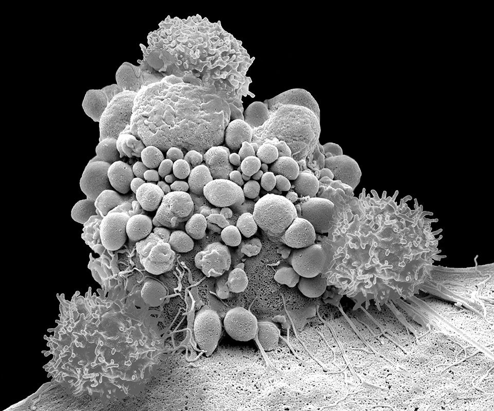
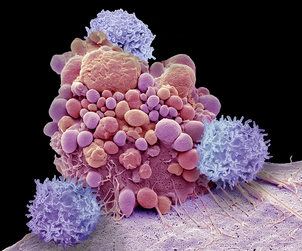
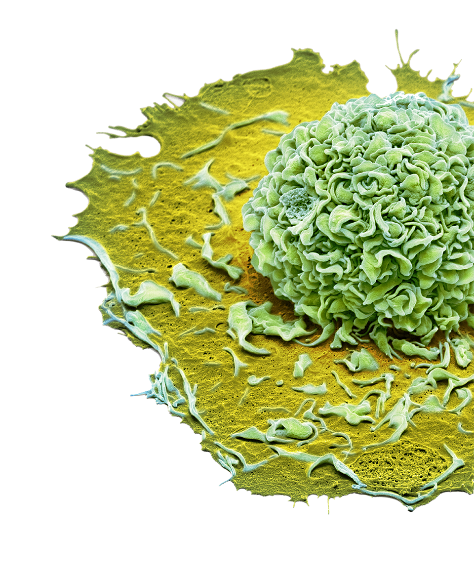
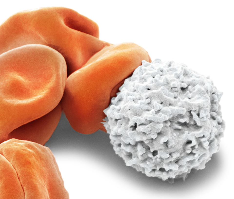
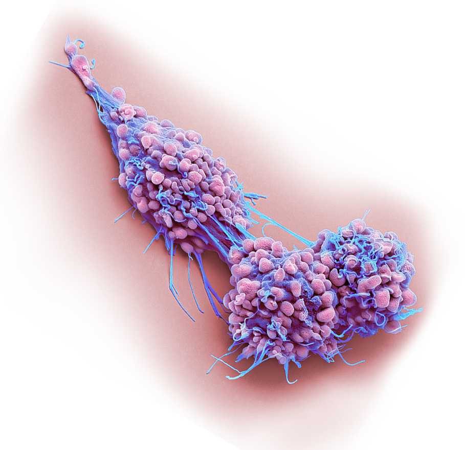
He trained at the Royal College of Surgeons and worked at Cancer Research UK. His coverage of the microscopic world is comprehensive and wide ranging, including bacteria, human and animal tissues such as blood, nerves, bone and hair cells, pests, parasites and pond life, electronic circuitry and nanotechnology. His greatest contribution is an unrivalled collection of difficult to access cancer cells.
SPL is the exclusive representative of his collection.
How and when did you become interested in science and microscopy?
I always had a love of natural history.
As a child I spent many hours bird watching, trawling rivers and ponds, and collecting anything interesting including stag beetles and slow worms.
After A levels in zoology and botany, I chose to pursue a degree in zoology.
What influenced you to go into this field?
I was given a great introduction to all forms of microscopy in my first job in the Anatomy department of the Royal College of Surgeons. It was my first encounter with the Electron Microscope and I worked on subjects such as leprosy, nerve repair and wound healing.
My next job in the Electron Microscopy department at Cancer Research UK gave me extensive experience in a whole range of cancer-related research subjects.
I still remember my first meeting with Rose, Creative Director at SPL, who introduced the idea that SEM images could be used creatively. That led me to pursue this idea seriously and a few years later I embarked on this journey full time with SPL.
How do you create an image?
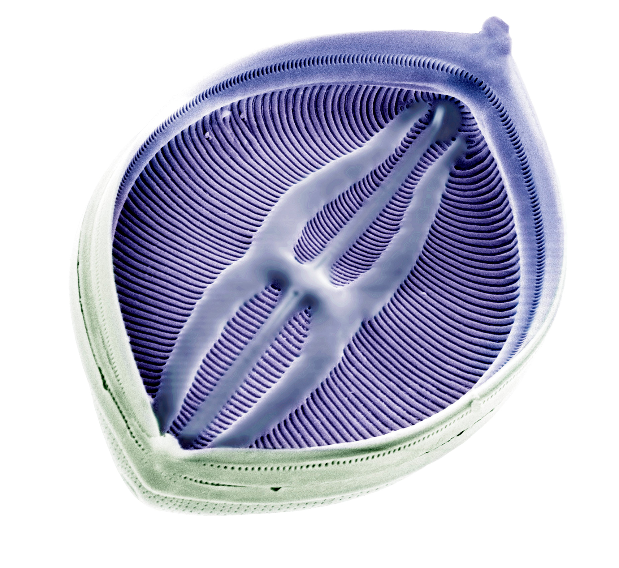
The scanning electron microscope (SEM) produces images by scanning the sample with a high-energy beam of electrons in a vacuum. As the electrons interact with the sample they produce secondary electrons, backscattered electrons and characteristic X-rays. These signals are collected by one or more detectors to form images that are displayed on a computer screen.
A sample is usually fixed (preserved), dehydrated and critical point dried in equipment that controls for distortion as the sample dries. This is an essential step in maintaining specimen quality.
The specimen is then coated in an extremely thin layer of heavy metal, usually gold, in order to reflect electrons and prevent specimen damage.
Could you explain how you colour the image?
An electron microscope can only produce images in black and white. Colour is a result of visible light. Using electrons rather than light allows the microscope to capture much finer detail, as electrons are smaller than light waves, but they cannot capture colour. The black and white images are coloured in Photoshop.
My colouring has improved with practice and software improvements, as has the resolution of microscopes and therefore image quality, which is far superior today than when I first started.
Colouring is a skill that is always improving, but essentially it is based on individual preference. It can be done functionally, highlighting different structures; naturalistically, imitating nature or artistically, meaning free range.
Why is it important to colour the images?
How long does the process take?
We live in a world of colour; we know our way around in it. By adding colour the image is made familiar, easier to understand, part of the human visual landscape.
Colouring can also add information to the image by identifying parts of it. It is expected of modern imagery as opposed to historic or archival material.
Time taken to colour depends on the complexity of the image; some are quick, for example a red blood cell, while others are hideously complicated such as cancer cells with multiple surface projections, dense networks of filaments and overlapping frills all bent on testing your masking skills. It can take an hour or a day. You need patience.
What’s your favourite imaging technique
SEM or TEM?
SEM because of the simplicity of preparation and the final 3D image.
How do you source your specimens?
Essentially anything that will fit in an SEM chamber, which is only a few centimetres wide, can be viewed.
I find specimens from diverse sources; collecting in the field, working with scientists and researching for suitable subjects. I’m no longer attached to an institution which means I beg, borrow and nag friends, family and strangers for samples.
I’ve been lucky recently to have established contact with a university professor keen to image some unique samples from a recent discovery. The sample was of unusual filamentous flu particles newly described by science. It was an exhilarating moment when they came into view under the microscope and it dawned on me I was the first person ever to see them.
My coverage is broad based. I’ll stick anything under the microscope to see what it reveals. Sometimes it is a waste of time and sometimes it takes my breath away at the sheer beauty and complexity. I like to be systematic in my attempt to cover as many tissues, cell types and natural samples as possible.
Do you consider your work art or science?
I’m really passionate about my work. Initially it was a scientific curiosity to understand and learn how biology works; to capture its structures and processes for the record. And only later did I begin to see with a more creative eye.
I’m often asked if the images I produce are art. If you define art as anything creative that causes emotion, then I believe my work combines science and art into one discipline. A lot of my images are used as standalone art and their diverse usage in the creative industry shows their artistic merit.
Personally, the distinction is not that important. What is important for me is that my images give me joy and pleasure, which is why I’m still creating at 71.
When was the first time you recognised your work was well received?
My first recognition came with winning the Visions of Science competition, where I received an overwhelming response to my work.
This was a nice surprise as I essentially work in isolation, on the microscope and when colouring.
My first scientific journal cover was also very exciting especially after 35 years of a scientific career.
Why do you think SEM imagery is so popular among the younger generation?
The unique 3D appearance means samples actually look more realistic than other imaging techniques, meaning people can relate more easily to the image.
SEM has the power to show complex scientific ideas in an understandable way and tell stories that children find fascinating and magical.
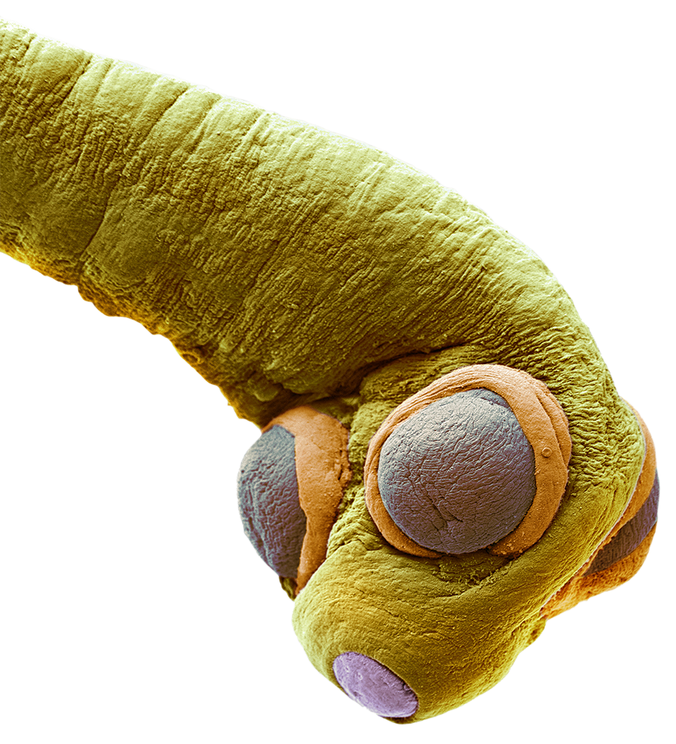
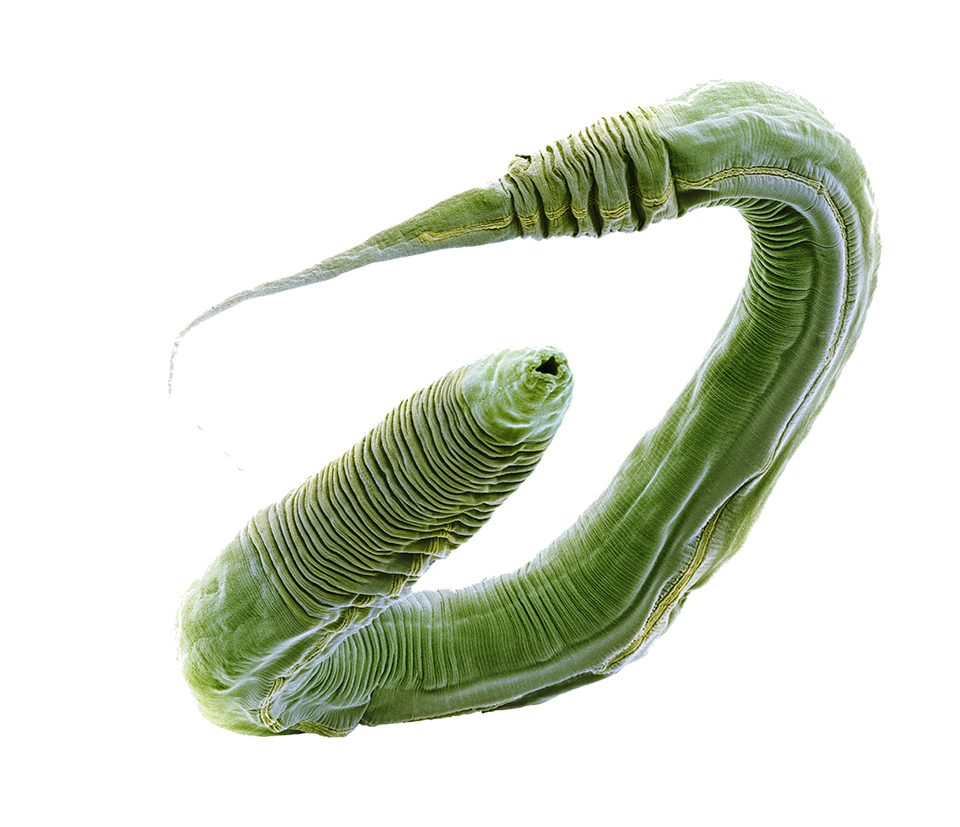
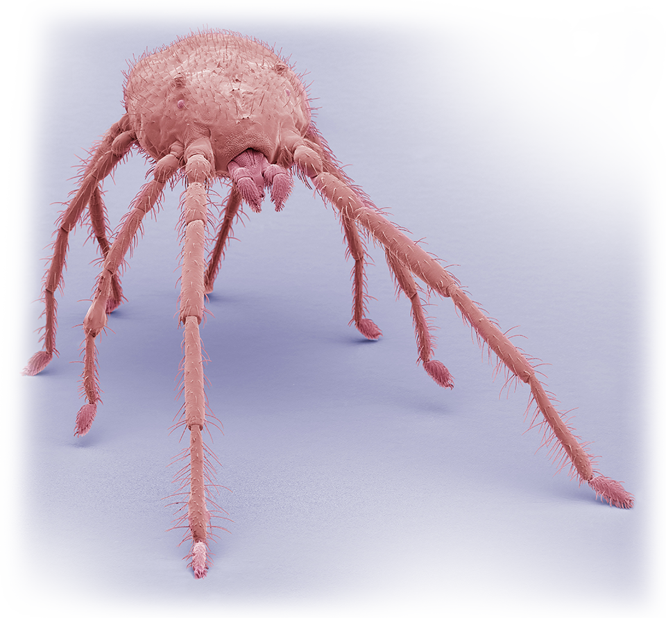
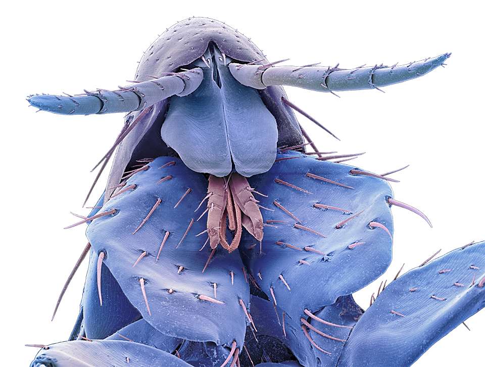
Invertebrates, especially insects, with their fine microscopic detail, are particularly popular and loved by children; the more bizarre and grotesque the image the better and louder the reaction. They like to be surprised, horrified and curious all at the same time.
Do you have any advice for younger microscopists?
Pursue your passion.
After nearly 50 years I am still seeing and taking unique images of things never captured before.
There is still a vast, microscopic world waiting for them to explore.
What is it about SEM that fascinates you?
Finding the unexpected.
Steve's
work in action


