Abstract Bodies

What initially drew you to the world of microscopy?

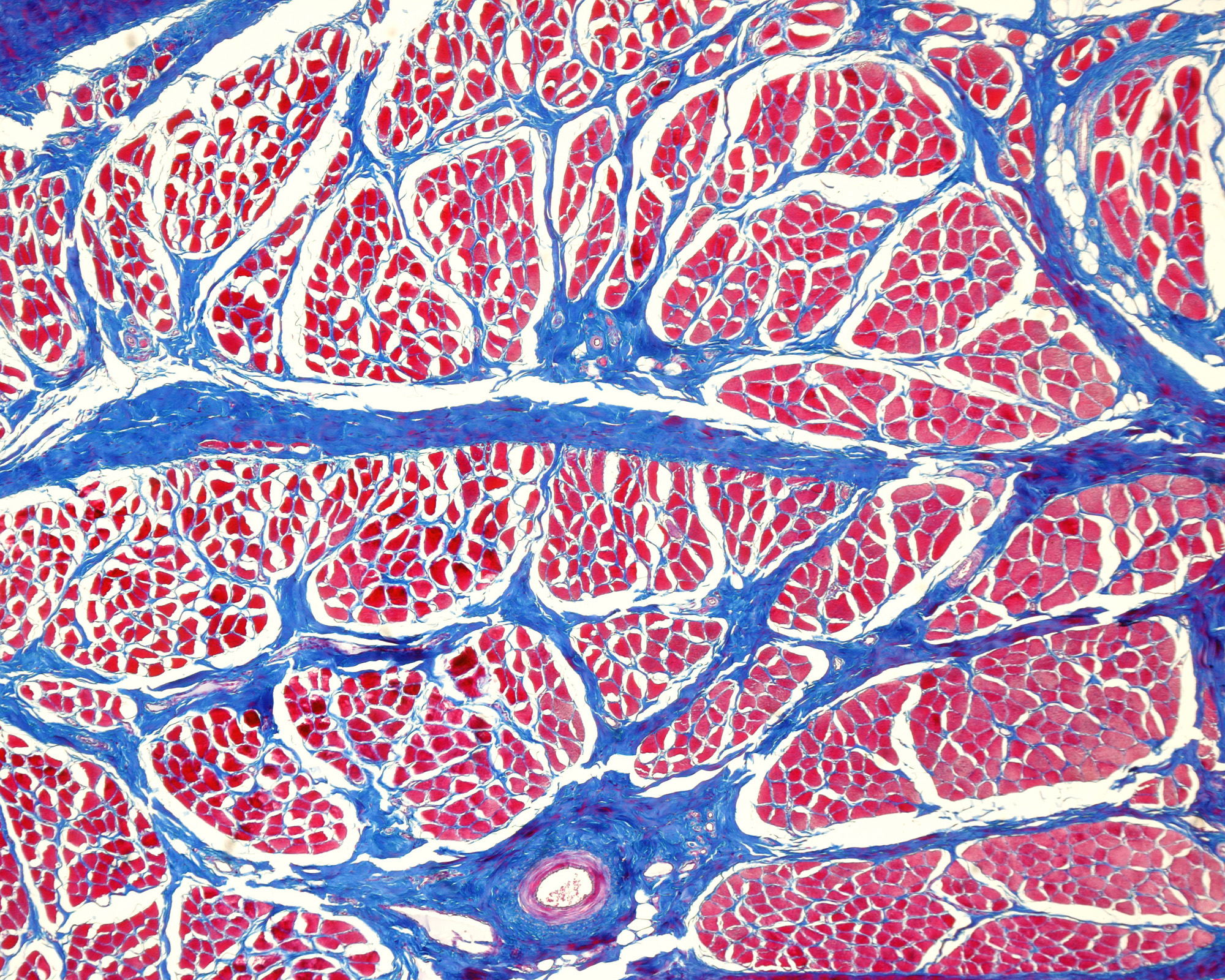
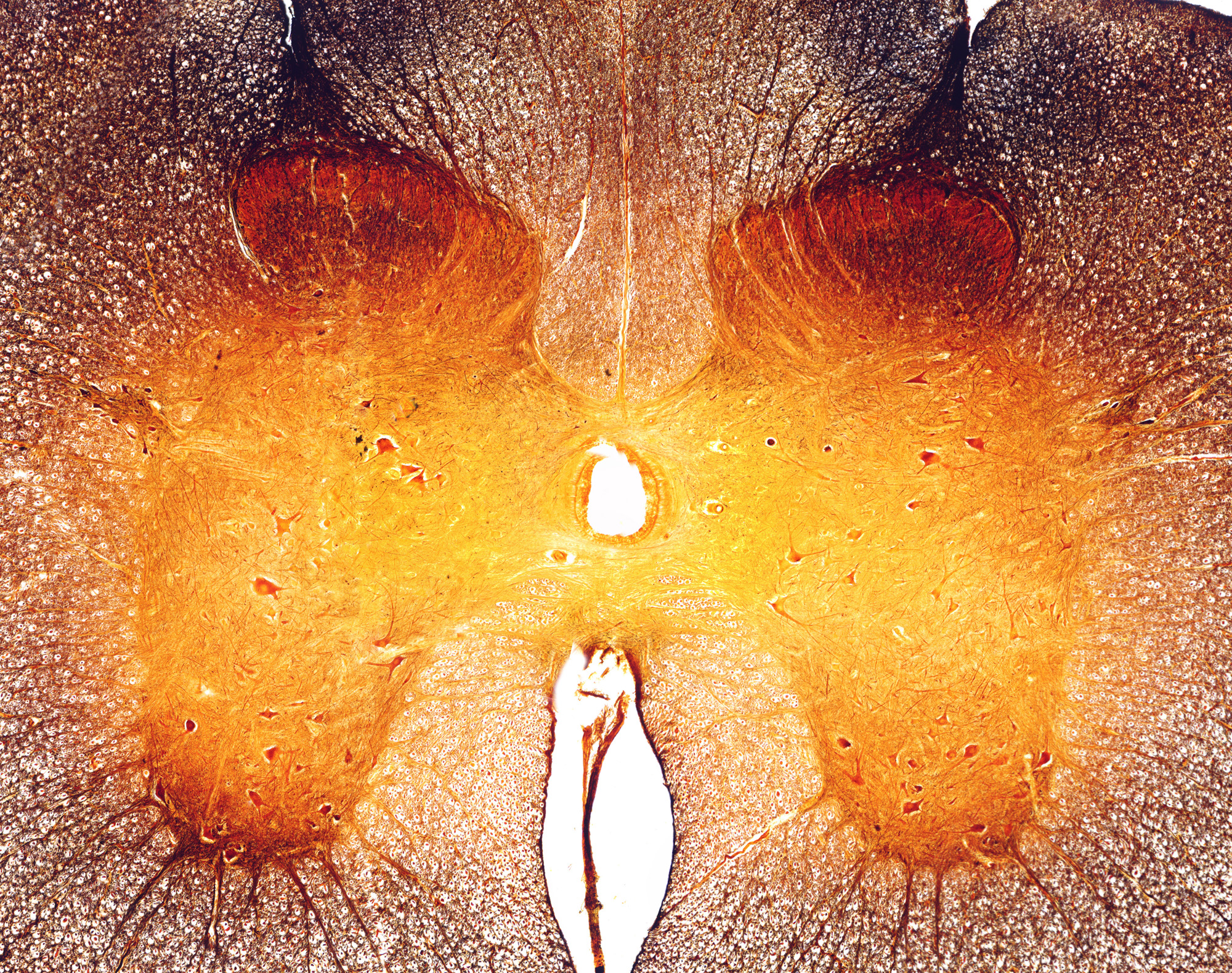
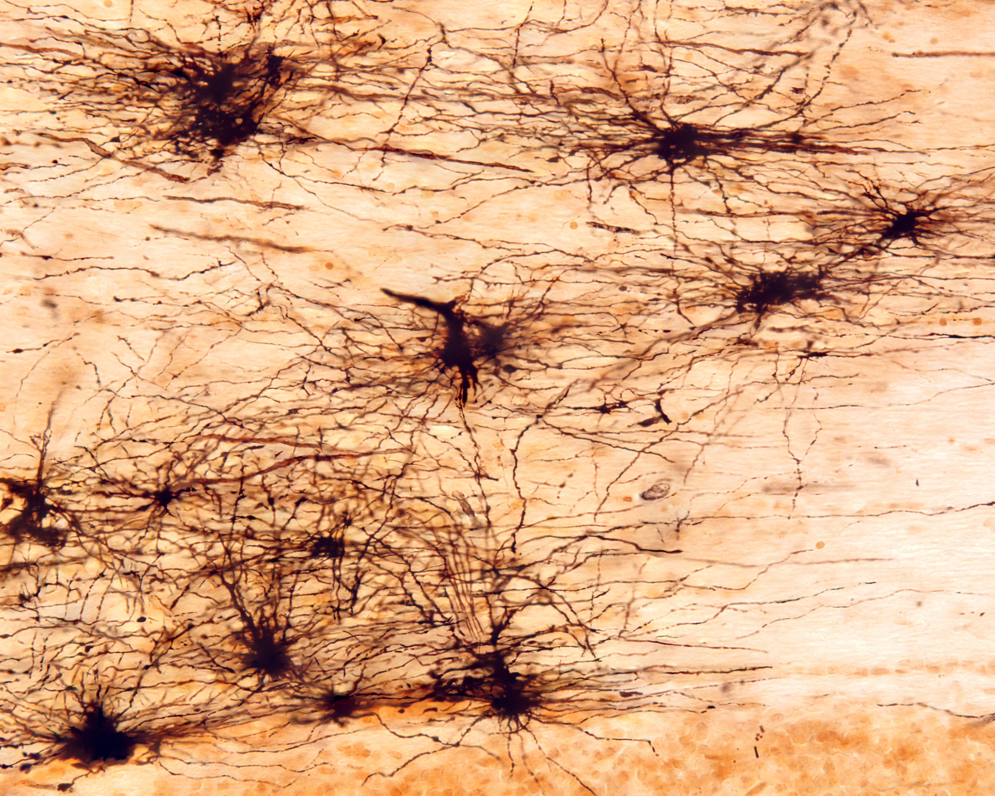
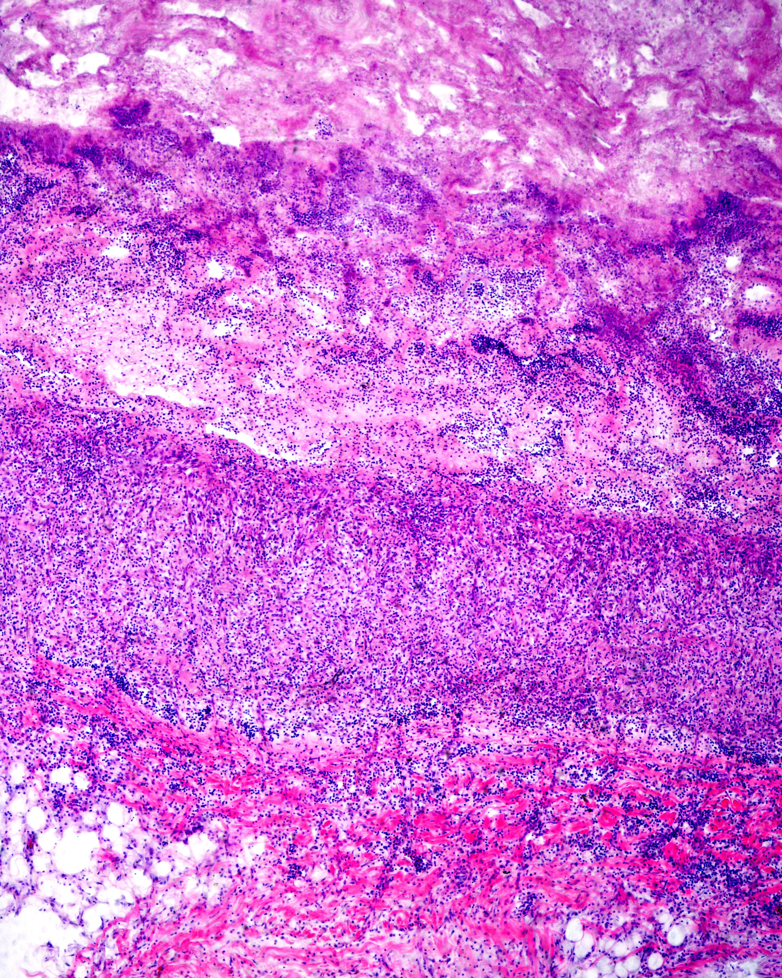
We have been attracted to the microscopic world since we were children. One of us got a microscope when he was only 10 years old, gifted by his parents.

We have always been passionate about the microscopic world, about how living things are made and how they work.

Our medical studies confirmed our interest in histology.

What challenges do you encounter when capturing light micrographs, and how do you overcome them?

The challenge is to obtain the perfect image, which is both demonstrative and visually attractive.

To do this, we first try to ensure that the histological preparation is sufficiently explanatory to be able to understand the structure being studied, looking for the most appropriate technique, in the most favourable organ, tissue and even species.

Secondly, the micrographic image must be as faithful as possible to how it looks under the microscope, to demonstrate as clearly as possible how the tissues of the organism are structured.

What is the importance of staining techniques in your images?

Histological technique in general and histological staining in particular are essential to achieve micrographic images of sufficient quality.

To obtain a good stain, it is essential that the sample is perfectly fixed, which is the first step of the histological technique. In most cases this involves placing it in a fixing liquid, which gradually penetrates from the outside into the inside until it is fully fixed. A poorly fixed sample will never achieve good staining results.

There are countless staining techniques, but a good stain with haematoxylin-eosin, the routine stain, shows many structures. Obviously, many other techniques are needed to demonstrate specific details.

How important are your images for medical diagnoses and scientific training?

There are numerous situations in which medical diagnosis and differential diagnosis are based on searching for a specific image. This includes not only microscopic images but also those obtained through ultrasound, radiology, or resonance imaging. Focusing on the microscopic world, we encounter everything related to biopsies, which are the essential tool for diagnosis, especially in tumour pathology.

In the scientific field, there is a need for a database of images that is as extensive, detailed, and rigorous as possible, showcasing normal tissue and cellular structures. This is essential for accurately assessing the responses to various experimentally induced conditions.

As teachers of Histology at Madrid University what is the most important lesson you try to convey to your students?

One aspect we have always emphasised in our teaching whenever possible is the relationship between structure and function. This approach helps students better understand the organisation and functioning of the human body through microscopy.

As for values, we focus on rigour and curiosity. Rigour is crucial because, in their future profession, incorrect or incomplete solutions will not be acceptable when treating patients. Curiosity is equally important, as it drives them to question their knowledge and to never accept anything solely on the basis of authority.

Would you agree there is an intersection between art and science in your work?

We look for certain aesthetics in order to better understand the concepts we want to convey.

An image must be demonstrative, but if it is also beautiful, it will be easier to assimilate. The artistic approach has a more creative component, where aesthetics is deliberately sought.

In our case, however, we prioritise the clarity of the image, adhering to the idea that a picture is worth a thousand words, over mere aesthetics. If, in addition to clarity, there is also an aesthetic component, all the better.

When did you first become aware that your images were both scientifically important and aesthetically pleasing?

We have had many favourable comments from our colleagues about the quality of our images in presentations, congresses and symposia. We have recently been featured on the cover of several scientific journals such as Endocrine Reviews and JCEM (5 of the 12 covers in the last year).

Of course, the positive reception of our micrographs by Science Photo Library is very important. We particularly appreciate the aesthetic value when people outside the field of histology are drawn to the images for their beauty, especially when artists view them and highlight their aesthetic qualities. We believe that the “artistic” element comes spontaneously because our first intention is to seek scientific value.

How has microscope technology evolved during your careers, and what impact has this had on your work?

A lot.

When we started, photomicroscopes used slides or film negatives. This was quite expensive due to the cost of film and the development process. As a result, there was a careful selection of what to photograph. Additionally, storing all these images required a lot of physical space.

With the arrival of new digital technologies, the first step we took was to scan all photographs. When we had a photomicroscope with a digital camera, we photographed all the tissues and organs that we had previously photographed using analogue methods. This involved a lot of work but brought significant benefits: improved image quality, easier focusing, and the freedom to take unlimited shots. In addition, the way of archiving and handling digital images is very convenient.

Regarding microscope technology, we have kept pace with the development of transmission and scanning electron microscopy, as well as histochemical and fluorescence techniques. We have incorporated all these technologies, which has allowed us to achieve greater detail and specificity when showing cellular and tissue structures.

What do you hope your legacy as microscopists and artists will be?

Our legacy must be more as microscopists than as artists.

The result of many years of teaching histology is reflected in our development of two Virtual Histology Practices. These are available online and free of charge. One is dedicated to animal tissues and another to the microscopic structure of organs.

Ultimately, we aim for the educational work we have done throughout our lives to be utilised by as many people as possible who are interested in histology. If we also attract those who appreciate the artistic and aesthetic aspects, so much the better.

As artists, our ideal would be to present colour micrographs that approach the beautiful and artistic drawings with which 19th-century microscopists illustrated their work.

Explore the work of

José Luis Calvo Martín and

José Enrique García-Mauriño Múzquiz