Steve Gschmeissner
Cancer Cell Death
Imagine peering into a microscopic battlefield, where fast-growing cancer cells face off against chemotherapy treatment.
Microscopist Steve Gschmeissner explores this topic in his most recent collection, offering a glimpse into the inner workings of cancer cell death.
Final Moments
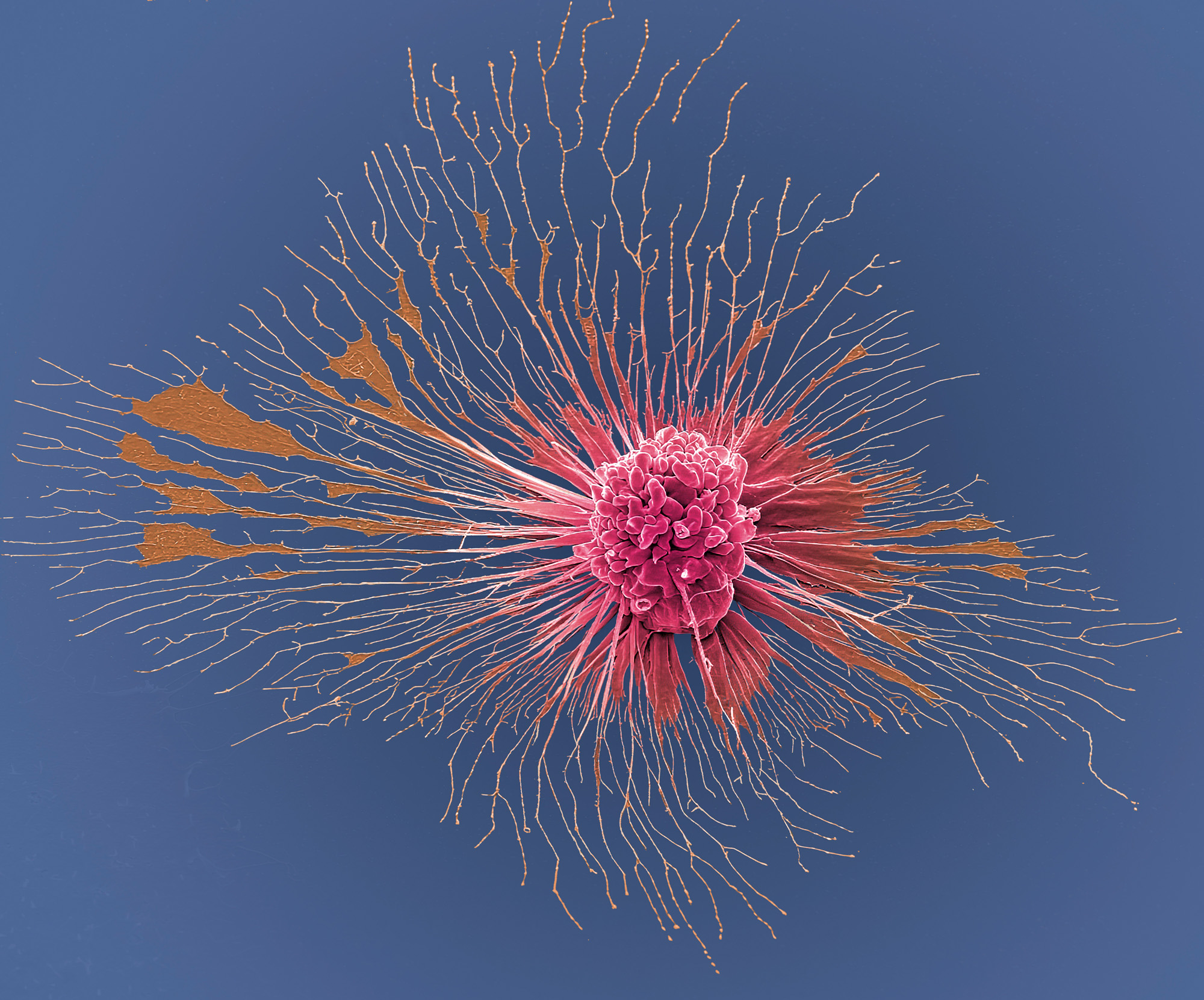
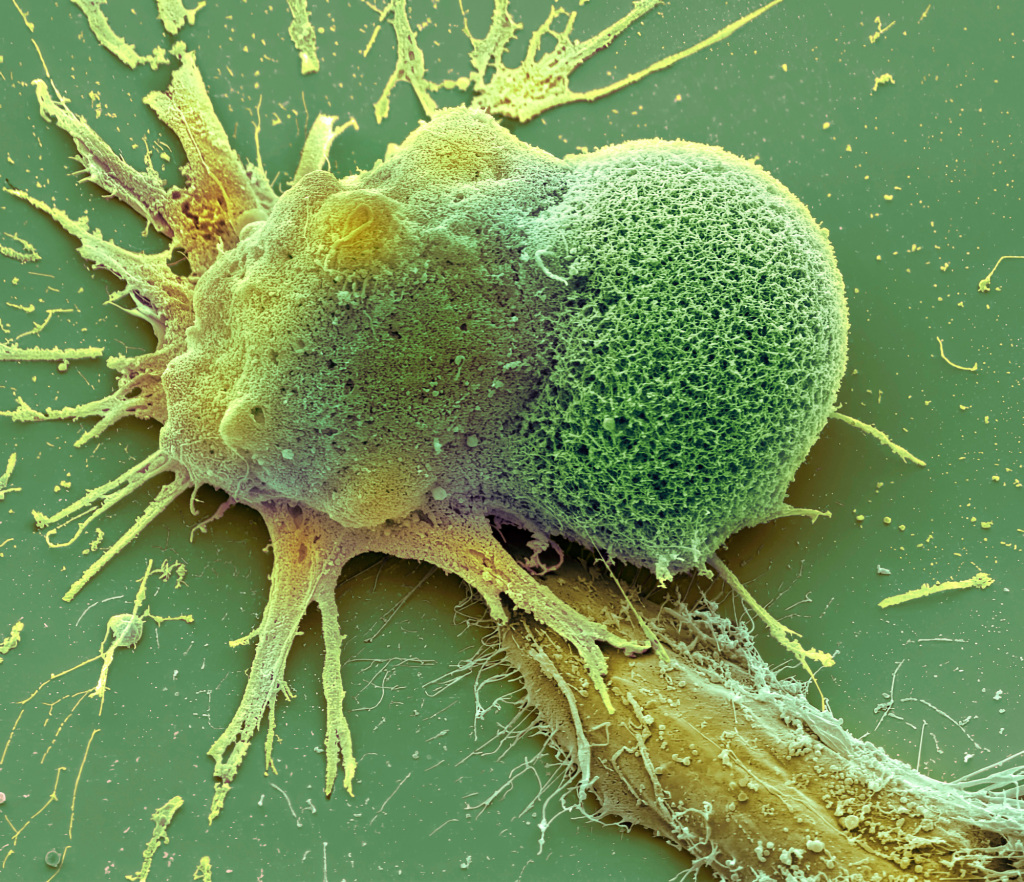
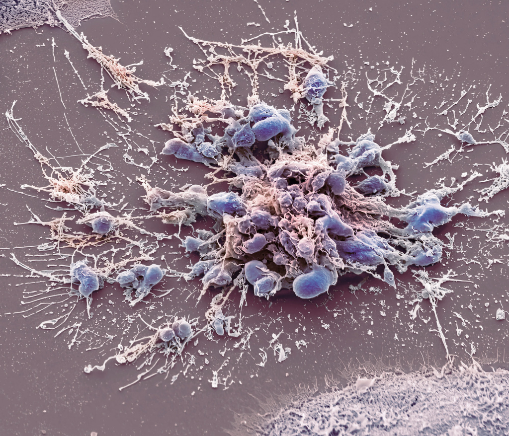
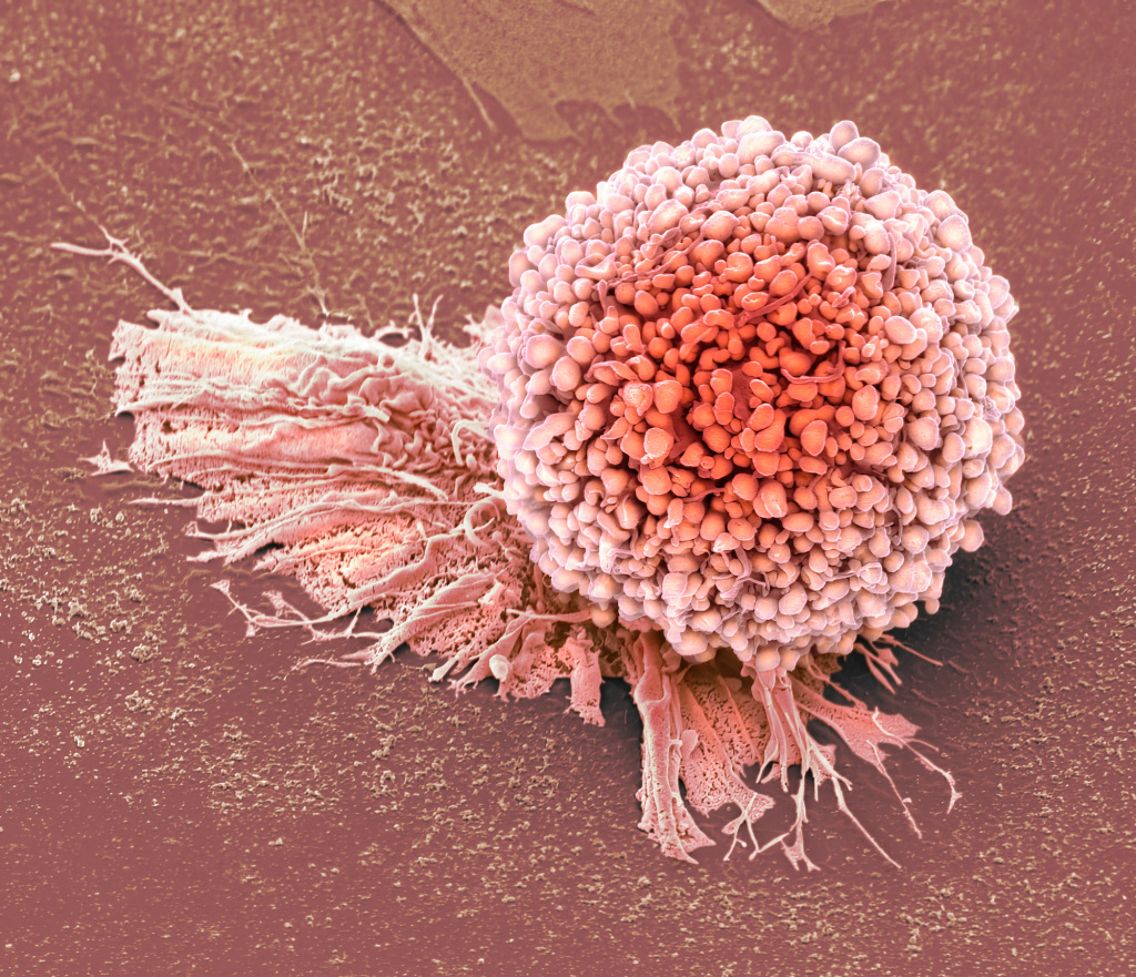
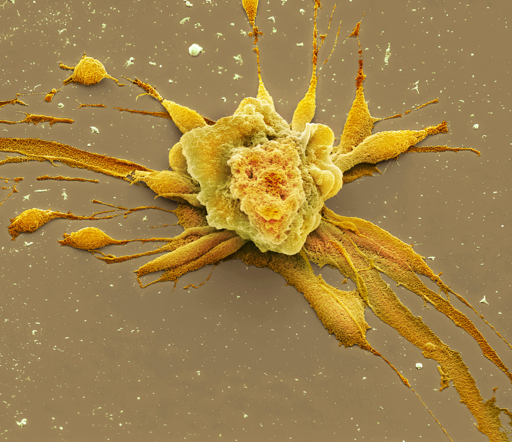
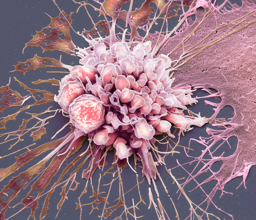
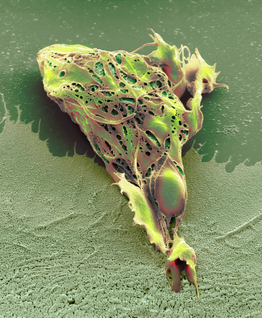
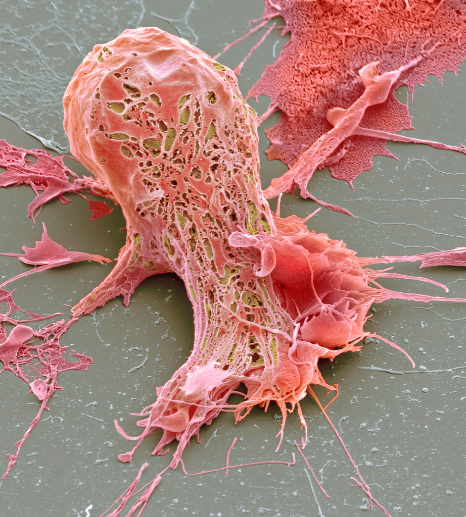
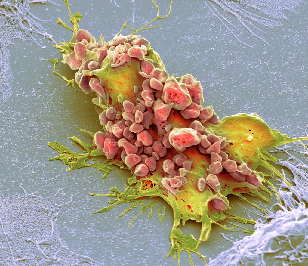
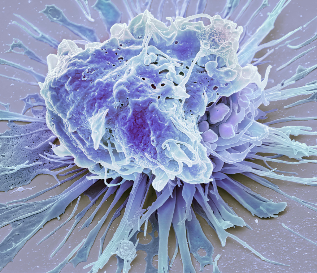
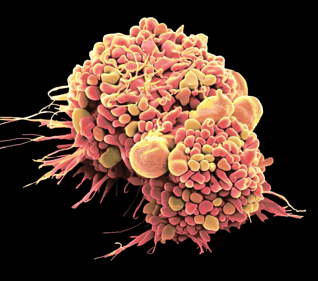
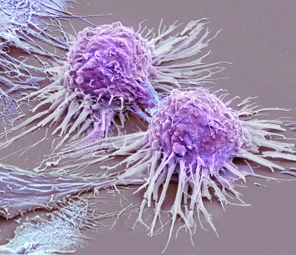
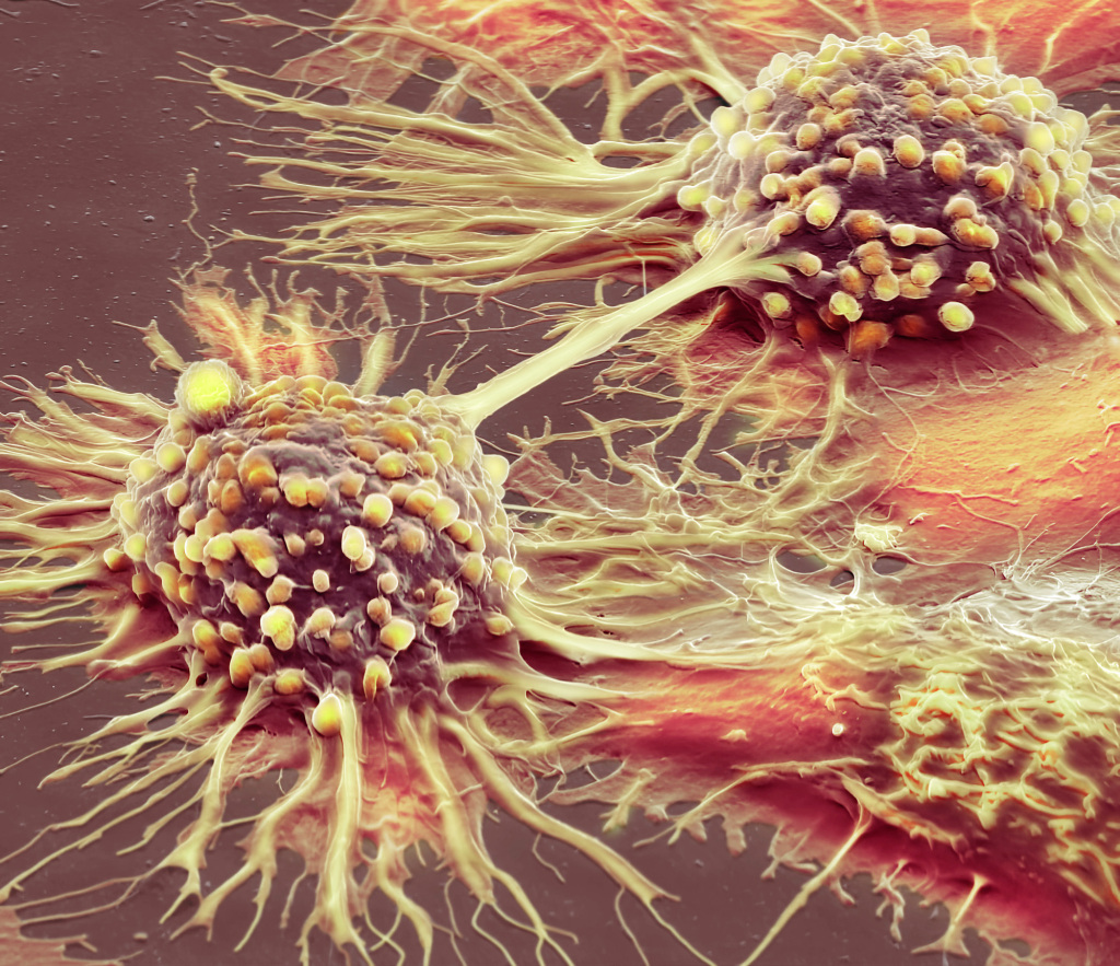
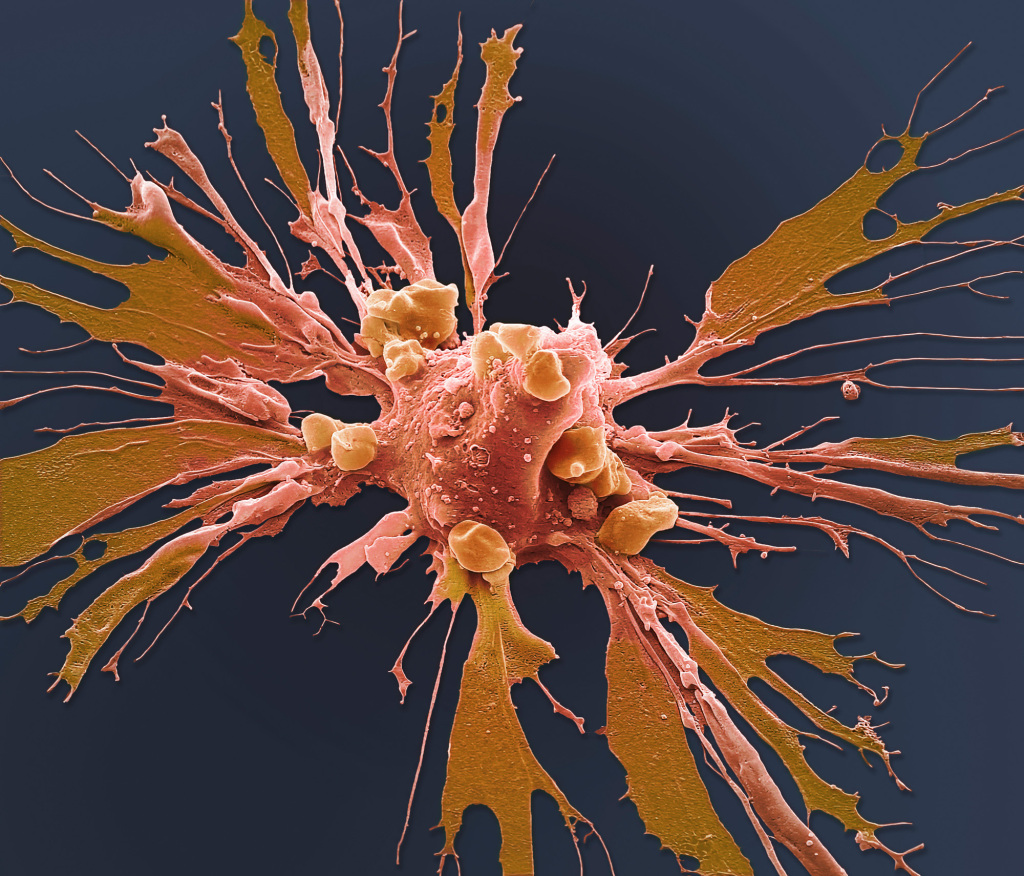
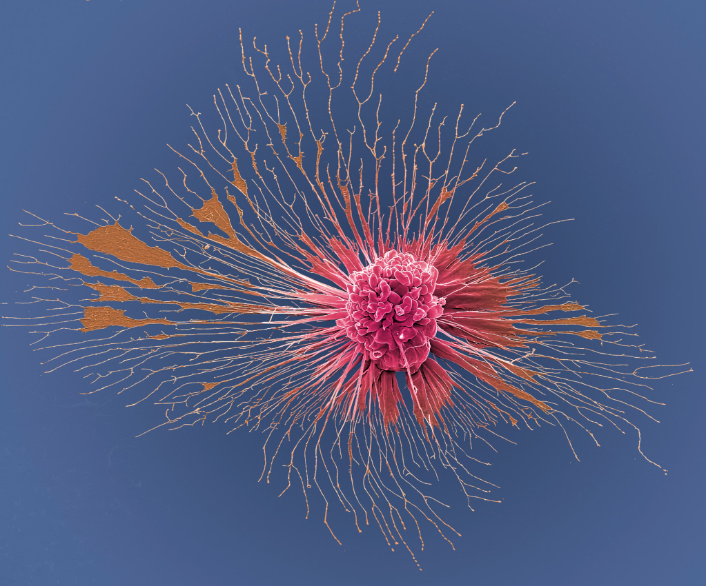
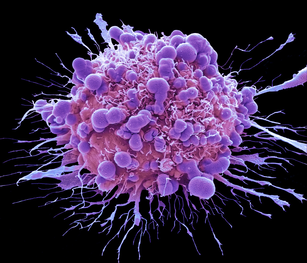
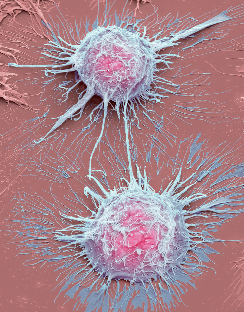
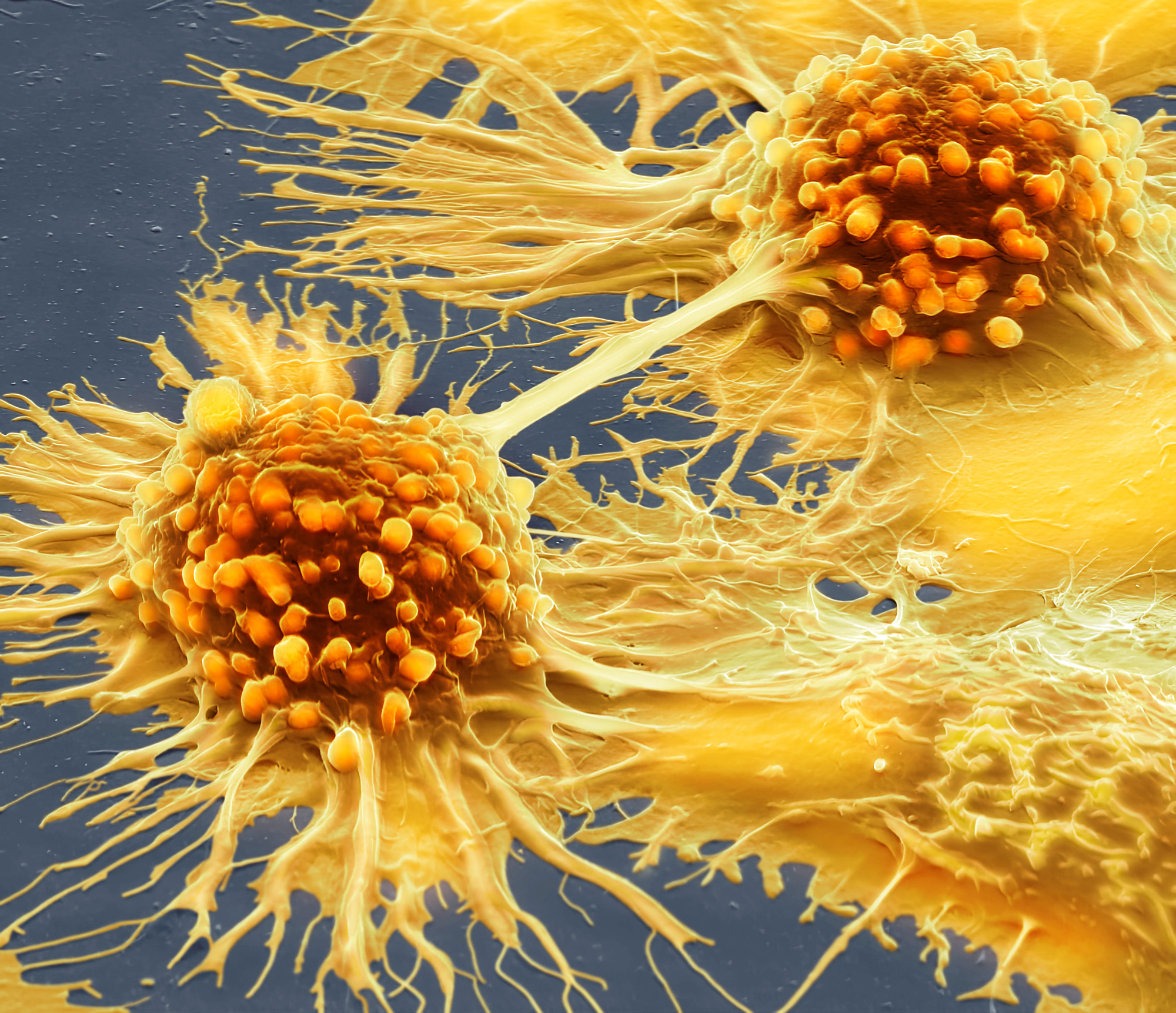
In this series of coloured scanning electron micrographs (SEMs), Steve Gschmeissner captures the final moments of cancer cells succumbing to chemotherapy.
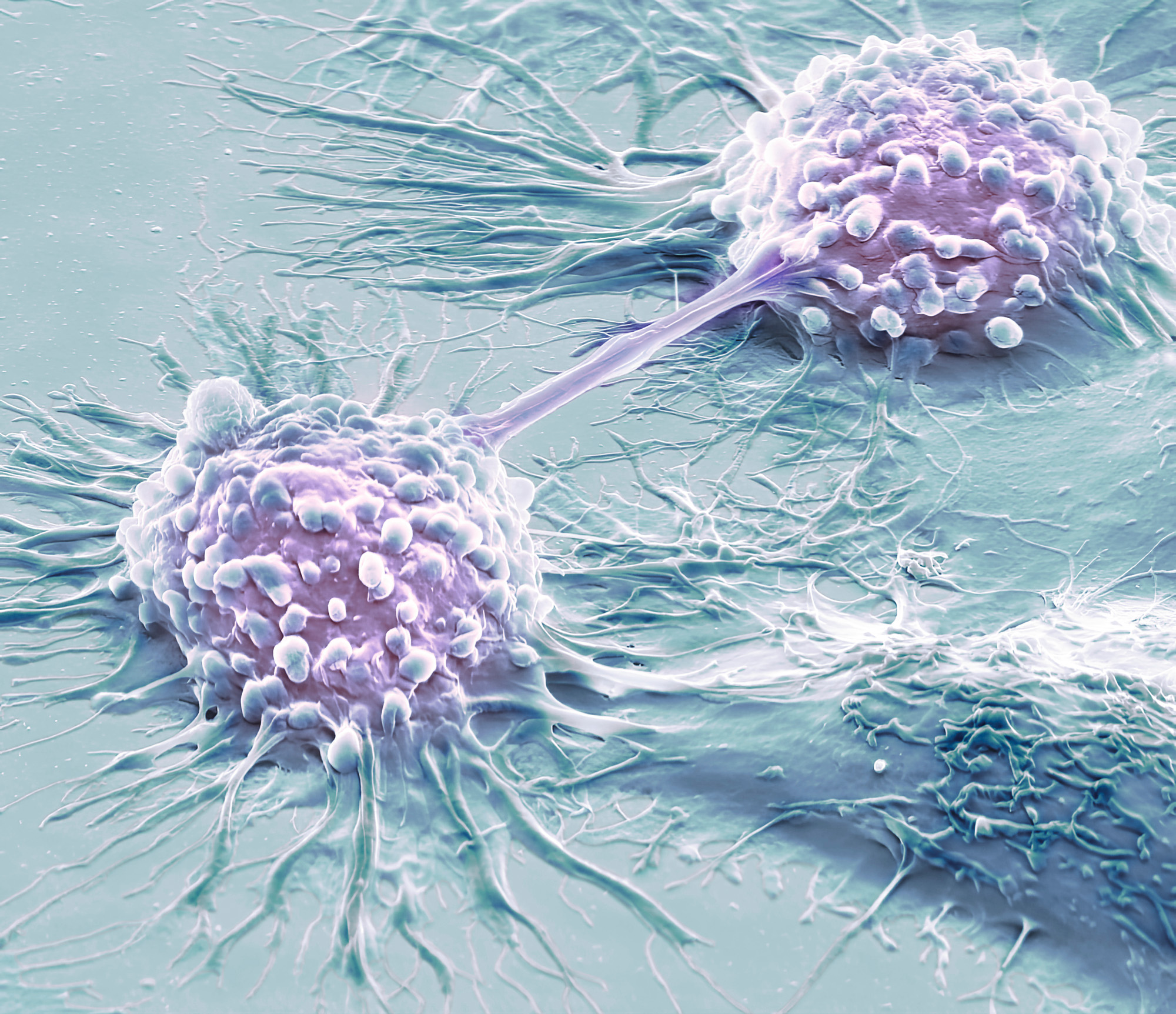
The project utilises HeLa cells to visualise cellular death triggered by the drug doxorubicin.
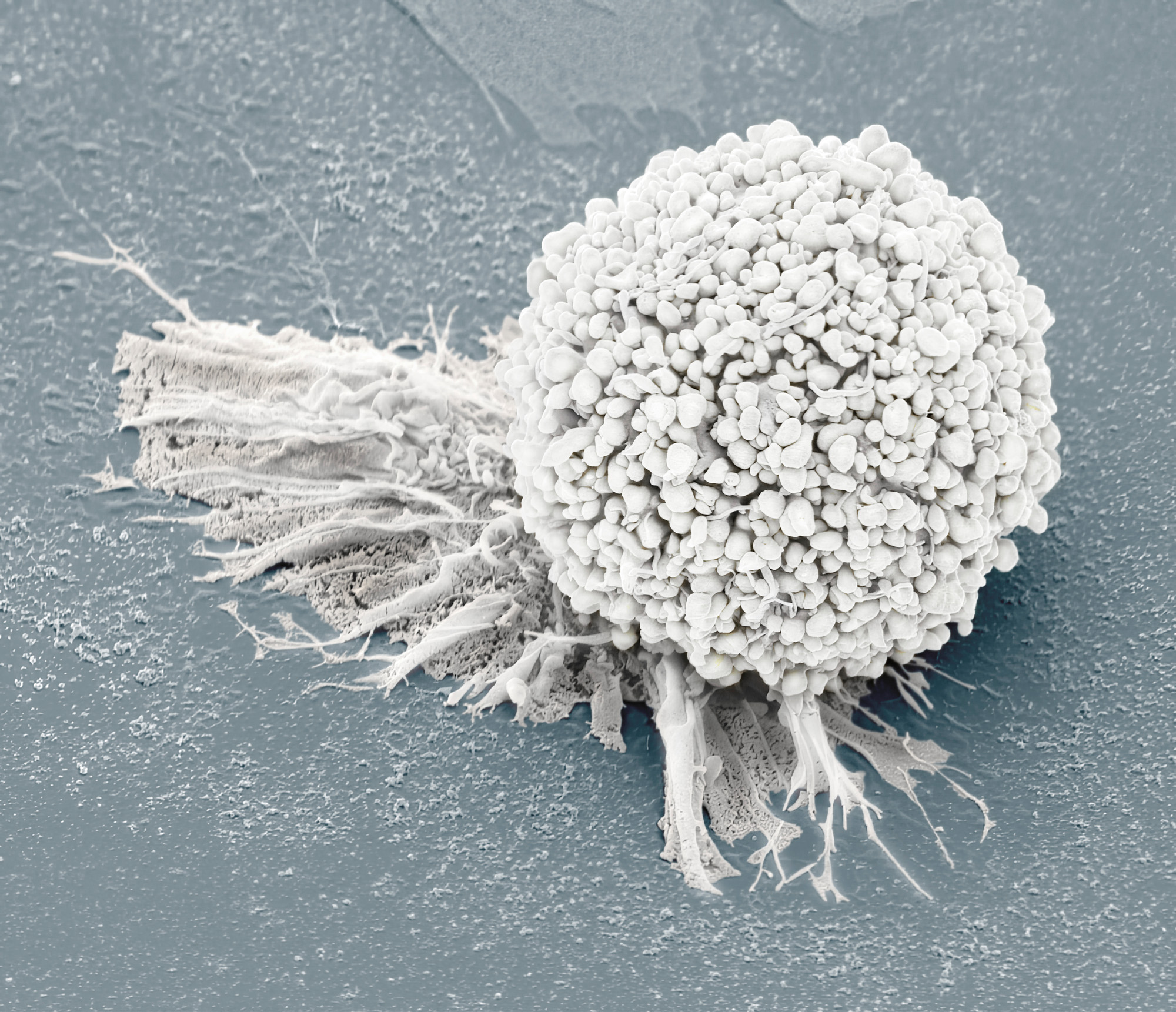

HeLa Cells
These cells are from a cell line taken from the cervical cancer of Henrietta Lacks (hence the acronym HeLa) in 1951 without her knowledge or permission.
Despite the ethical complexities surrounding their acquisition, Lacks’s legacy lives on in countless scientific advancements, including the fight against cancer.
Her unknowing contribution to science has also sparked important conversations about informed consent and racial inequities in healthcare.
Doxorubicin
Steve Gschmeissner’s collection focuses on the effects of doxorubicin, a powerful chemotherapy drug, on HeLa cells.
Doxorubicin works, in part, by interfering with DNA replication, targeting the much higher rate of cell division in tumours.
This disruption ultimately leads to cell death. However, the death of a cancer cell isn’t always uniform and this complex anticancer activity is still not fully understood.
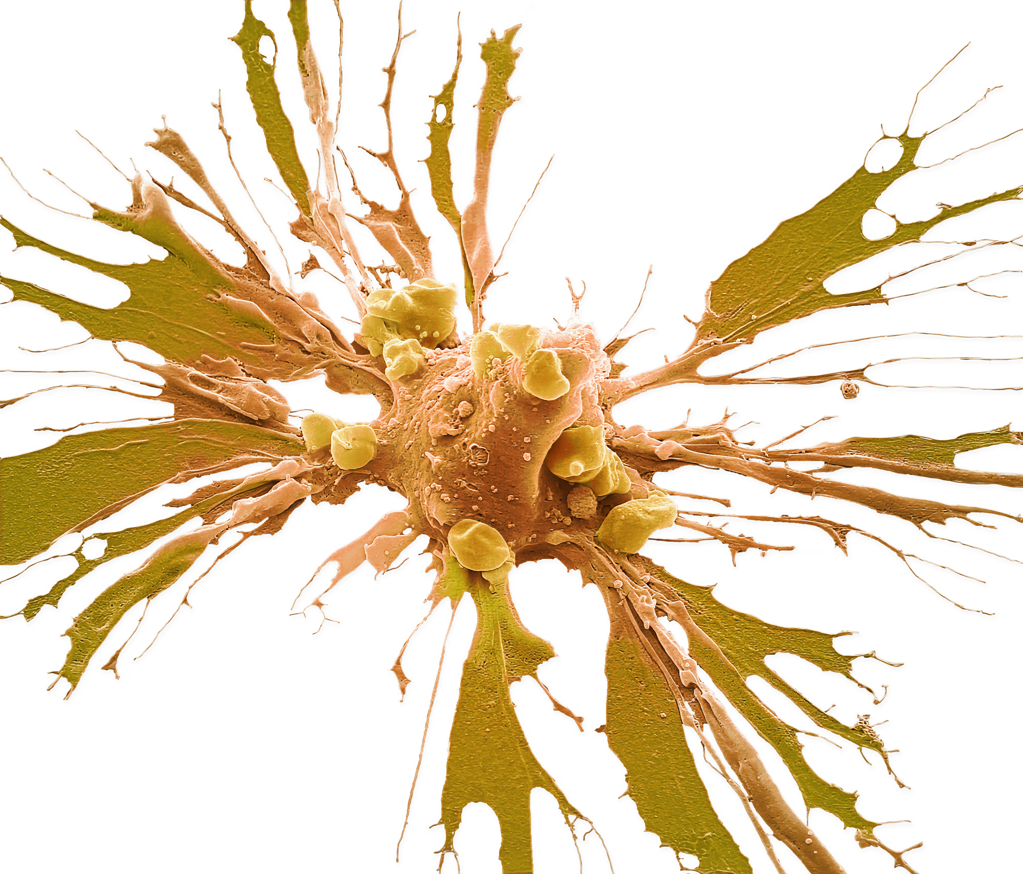
The two main pathways to cell death are apoptosis and necrosis.
Apoptosis
Apoptosis is a normal, ongoing process in the body. It rids the body of old, damaged cells, allowing younger, healthier cells to replace them.
When doxorubicin strikes, cancer cells initiate a self-destruct sequence (seen as blebs on the surface) causing the nucleus to shrink and fragment, destroying the cells in a controlled manner.
This demise mechanism minimises damage to surrounding tissues, making it a desirable outcome in cancer treatment.
Necrosis
Necrosis in contrast is an uncontrolled destructive process.
When the cell is overwhelmed by the toxic effects of doxorubicin its membrane, peppered with holes, finally ruptures, spilling its contents into the surrounding environment.
This method of death can trigger inflammation and harm healthy cells, which is why scientists strive to tip the balance towards apoptosis.
The Final Moment
Steve Gschmeissner is a renowned microscopist with a unique specialisation in capturing the unseen world of science.
His work is not only aesthetically pleasing; it also serves as a valuable tool for scientific communication. His images, often used in research papers and educational materials, bring scientific concepts to life in a strikingly beautiful way, inspiring awe at the sheer complexity of a teeming universe invisible to the naked eye.
This collection of images would not be possible without the invaluable collaboration of Professor Greg Towers at University College London.
