An award winning scientific photographer specialising in microscope art. Be mesmerised by the exquisite beauty and intricate patterns of crystals in nature.
Karl Gaff
A fusion of science and art
When did your interest in microscopy begin? Was it a childhood dream or something you discovered later?
I was probably about eight years old when I received my first microscope, albeit a toy, I was mesmerised by what I could see. I spent my childhood immersed in nature with my magnifying glass looking at plants and their flowers and collecting pond samples to bring home to look at them under the microscope.
The microscope was incapable of having a camera adapted to it at the time. Of course, as I look back, it would have also been far from capable of producing the dazzling results that I am used to today.
Over the years I slowly upgraded to better microscopes with the dream of someday becoming a professional microscopist and scientific photographer. It was that first experience back in 1992 that projected me on a trajectory towards a career and passion for science and a love of nature.
Could you give us some background on your experience and training?
I enrolled at the Dublin Institute of Technology in 2004, graduating in 2007 with a BSc in Physics & Life Sciences and again in 2009 with a BSc in Physics & Physics Technology. Following these, I undertook an MSc in Imaging & Microscopy in 2011 at University College Dublin (UCD).
I was employed by UCD as a microscopist in the Conway Institute of Biomolecular and Biomedical Research. I gained work experience on experimental design and operation of confocal microscopes for live-cell imaging, tissue preparation for transmission and scanning electron microscopy and image processing.
For the past 7 years I have worked at TU Dublin (formerly Dublin Institute of Technology) as a technical officer, managing the advanced undergraduate laboratories for final year physics. This involves the design, construction and testing of apparatus for experimental physics and supervising students in the final year of their physics degrees.
You’re particularly interested in crystallography. What is it about this technique that holds your fascination?
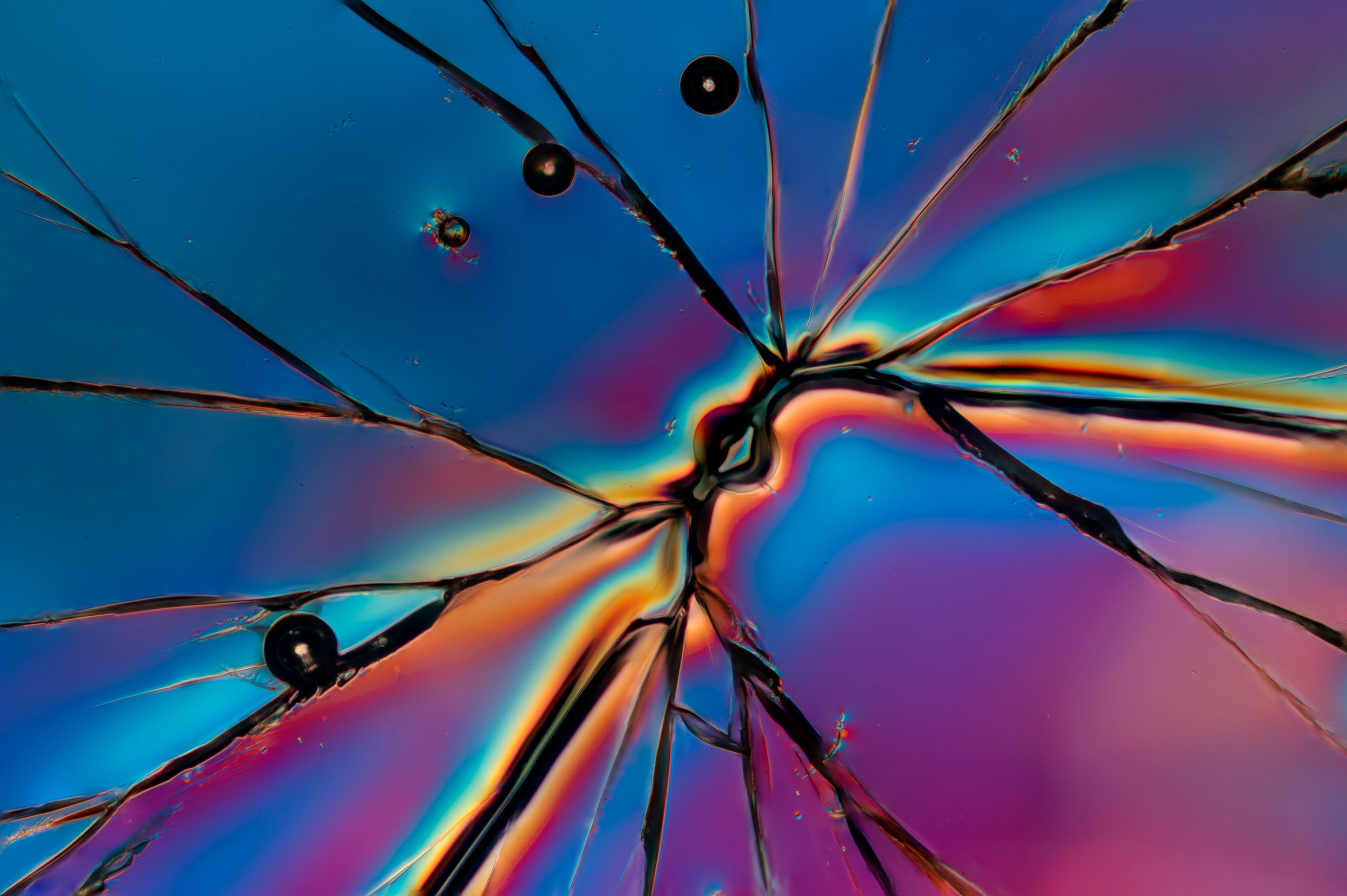
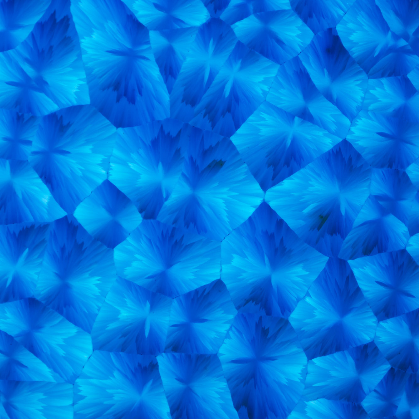
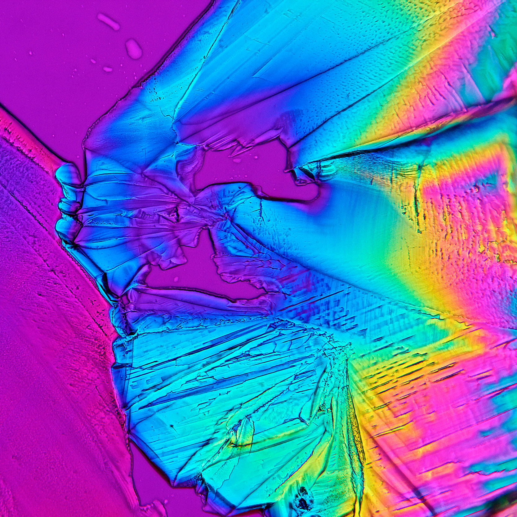
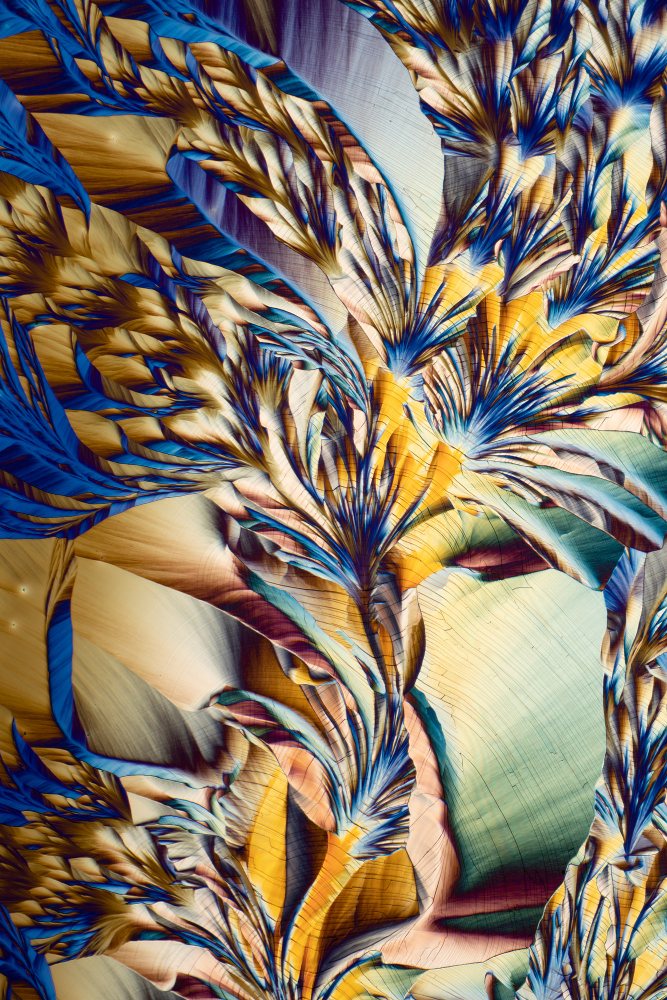
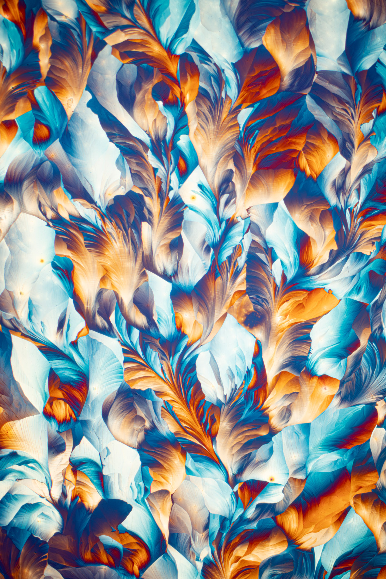
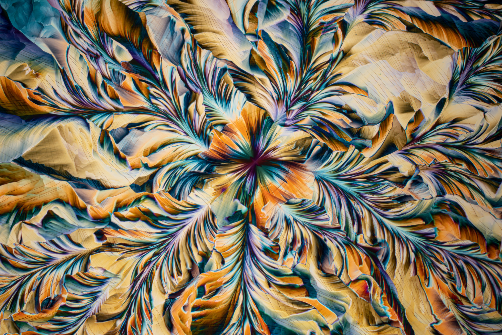
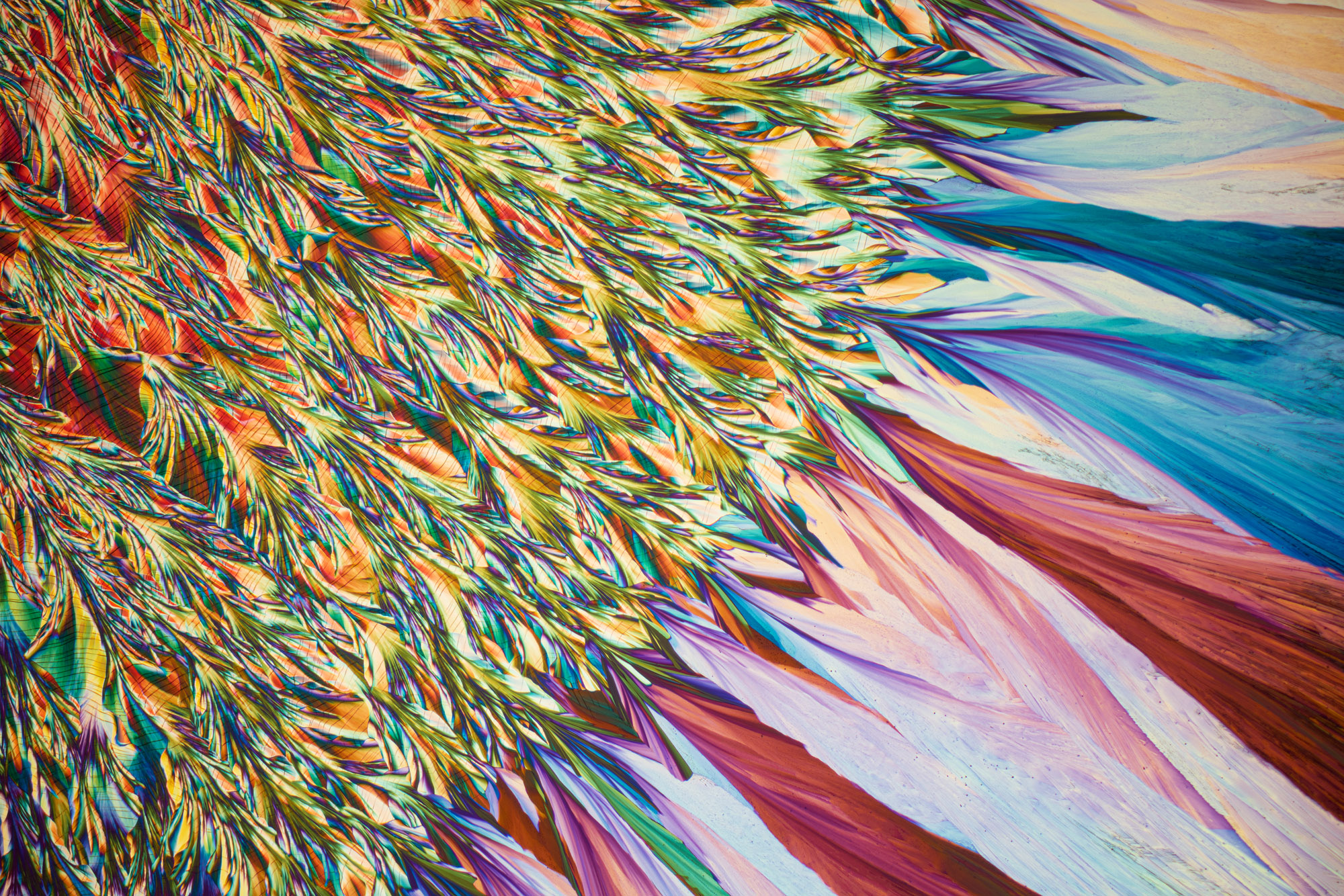
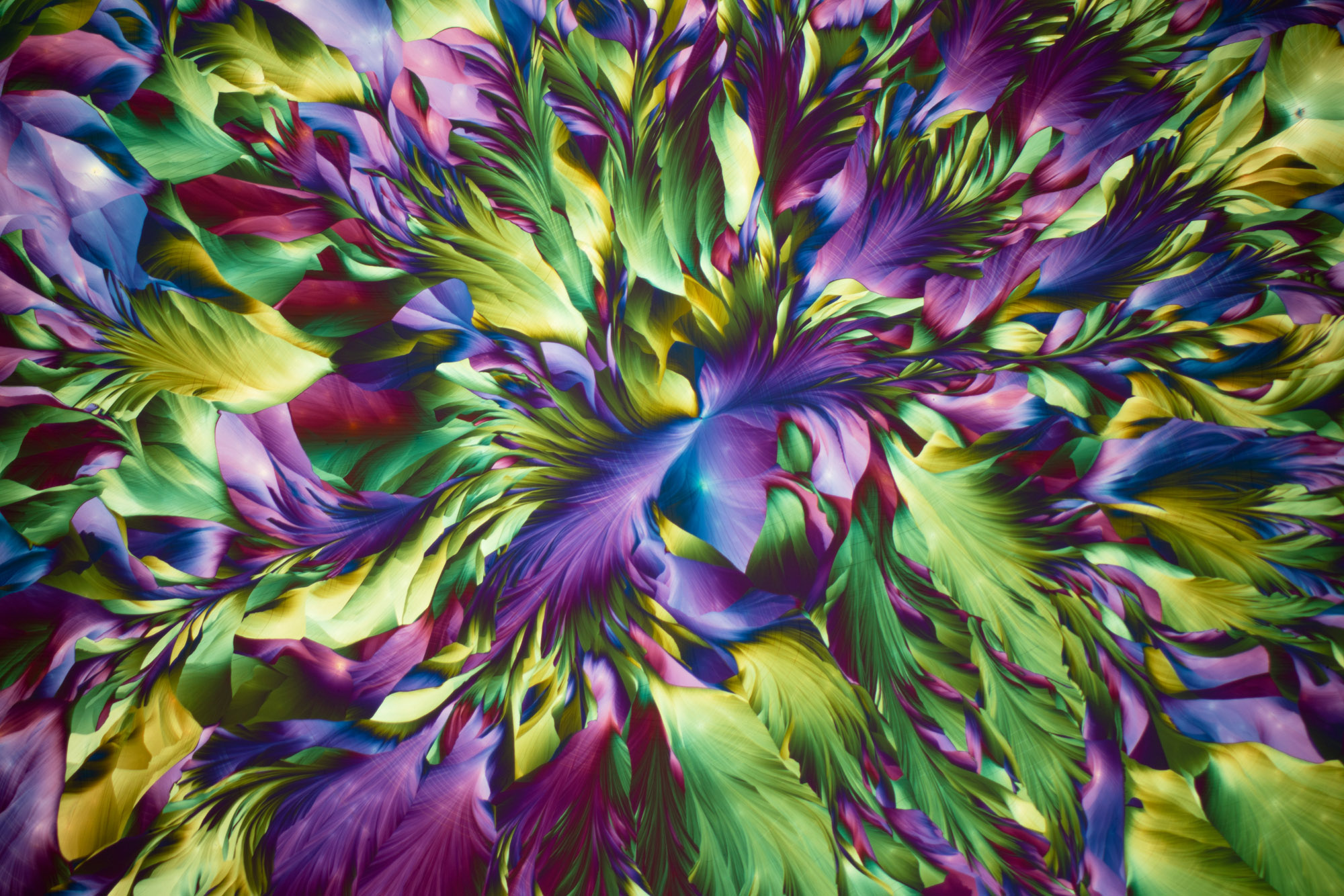
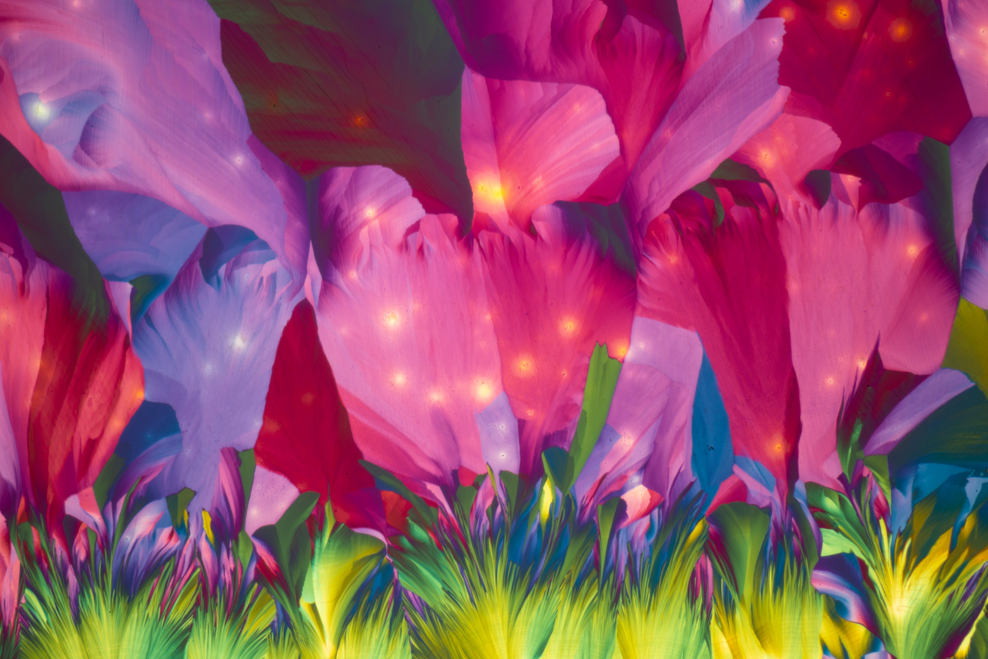
What fascinates me about imaging crystallised chemicals is the infinite variety of texture, pattern and form. You never know what to expect!
There are a multitude of factors at work in the system, some of which you have no control over including temperature fluctuations, thermal perturbations, humidity, sample impurities and the inherent randomness – they are truly complex systems and so because of this you never get exactly the same results as before. Slight changes in ratio of the mixture can also greatly alter the results you get.
It’s a research and discovery process. Sometimes you are disappointed and other times you are completely and utterly mesmerised by the views through the binoculars of the microscope.
How are the spectacular colours and shapes created? Do you have any control over them?
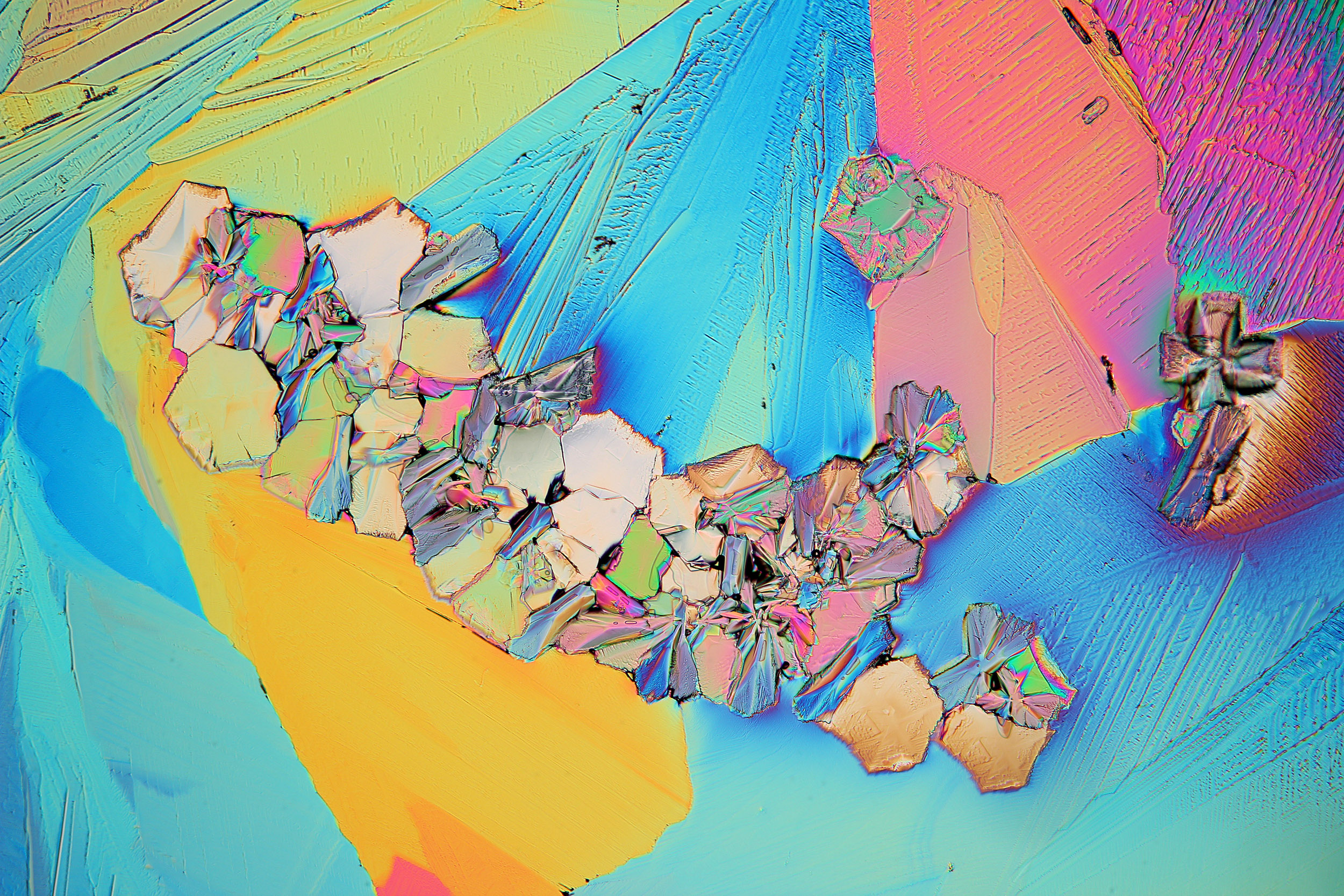
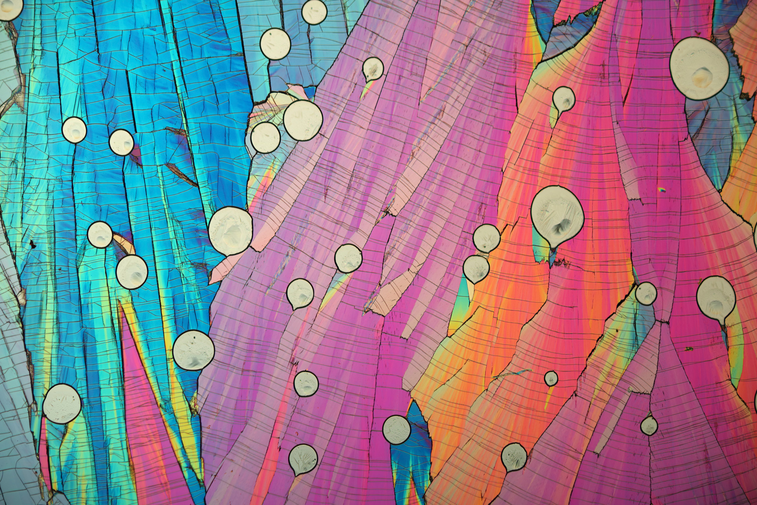
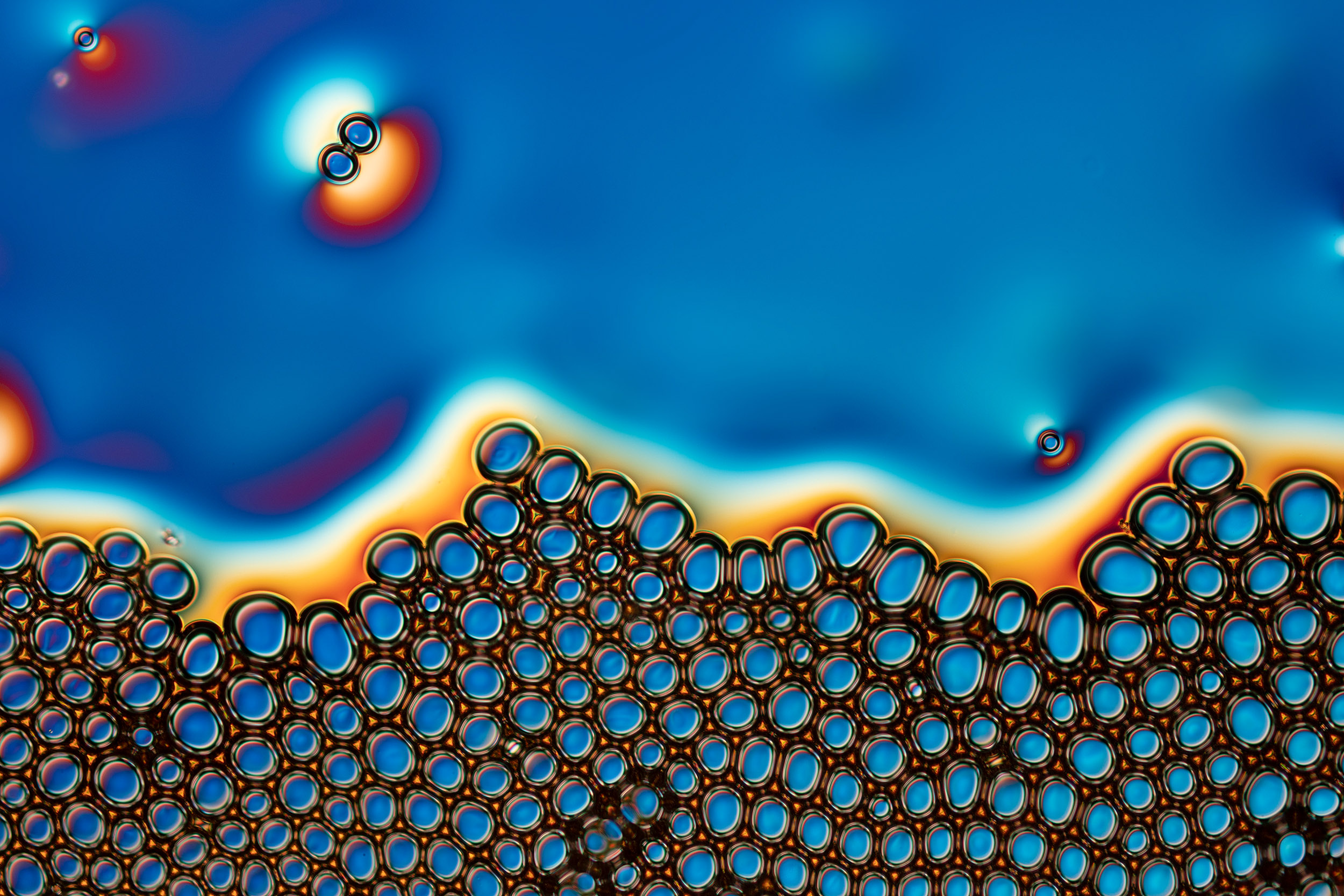
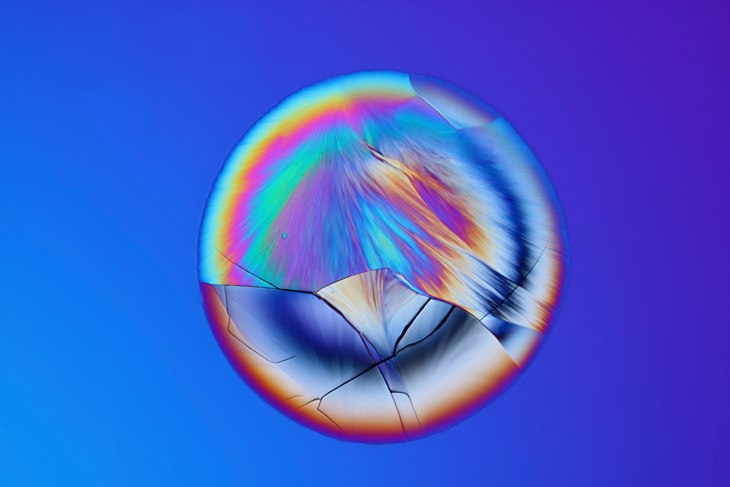
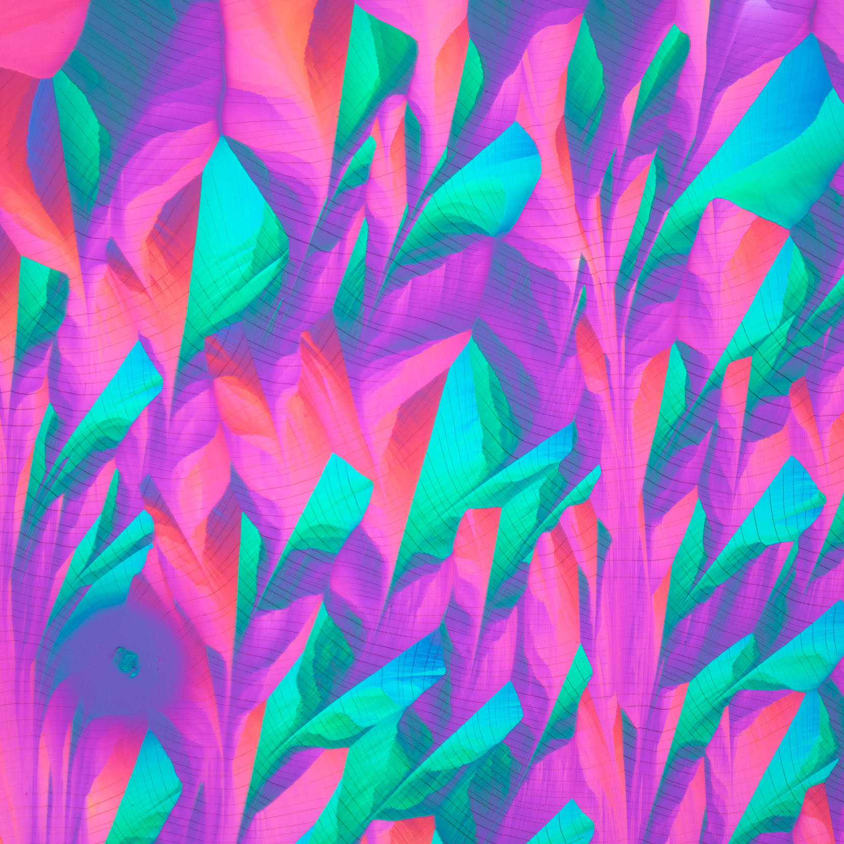
When the microscope is properly configured for compensated polarised light and the sample inserted onto the stage, Nature will choose her colour palette depending on the physics and chemistry of the sample.
There is limited control of the colours. When certain elements in the optical system are adjusted, different colours can be seen, depending on how the molecules in the sample manipulate the light as it passes through.
Molecules come in an infinite variety of shapes that link together like tiny magnets to form, in this case, crystals. The crystals are not pure however because the samples are dissolved in tap water which contains lots of different impurities like calcium, fluorine, chlorine and other metals. These impurities change how the sample molecules connect together and as a result causes the shapes to take on sometimes drastically different forms than if pure sample were used.
As is always the case, the overall structural formation of the crystallised sample can be thought of as being ‘kaleidoscopic’ in nature and it is this structure that dictates the coloured patterns that can be seen.
What are the challenges you encounter when producing images and videos?
All imaging is carried out in my home studio and so I am limited by the equipment that I have available to me. This is a challenge because I have many ideas that I would like to undertake that are not possible with the equipment that I have.
For example, some crystals form at such a fast rate that a high-speed camera is required to capture them and these cameras are extremely expensive.
You’re using such a diverse range of specimens. What triggers your decision and how does it affect the creative process?
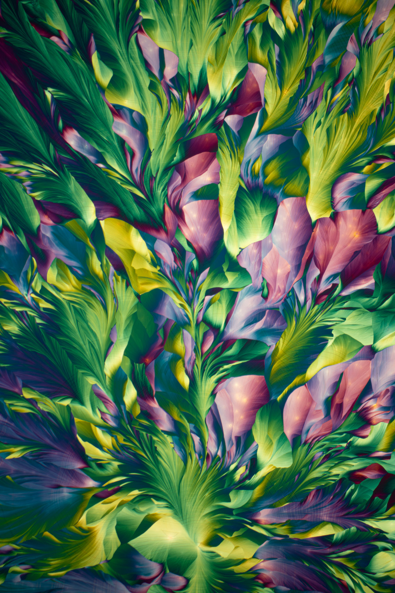
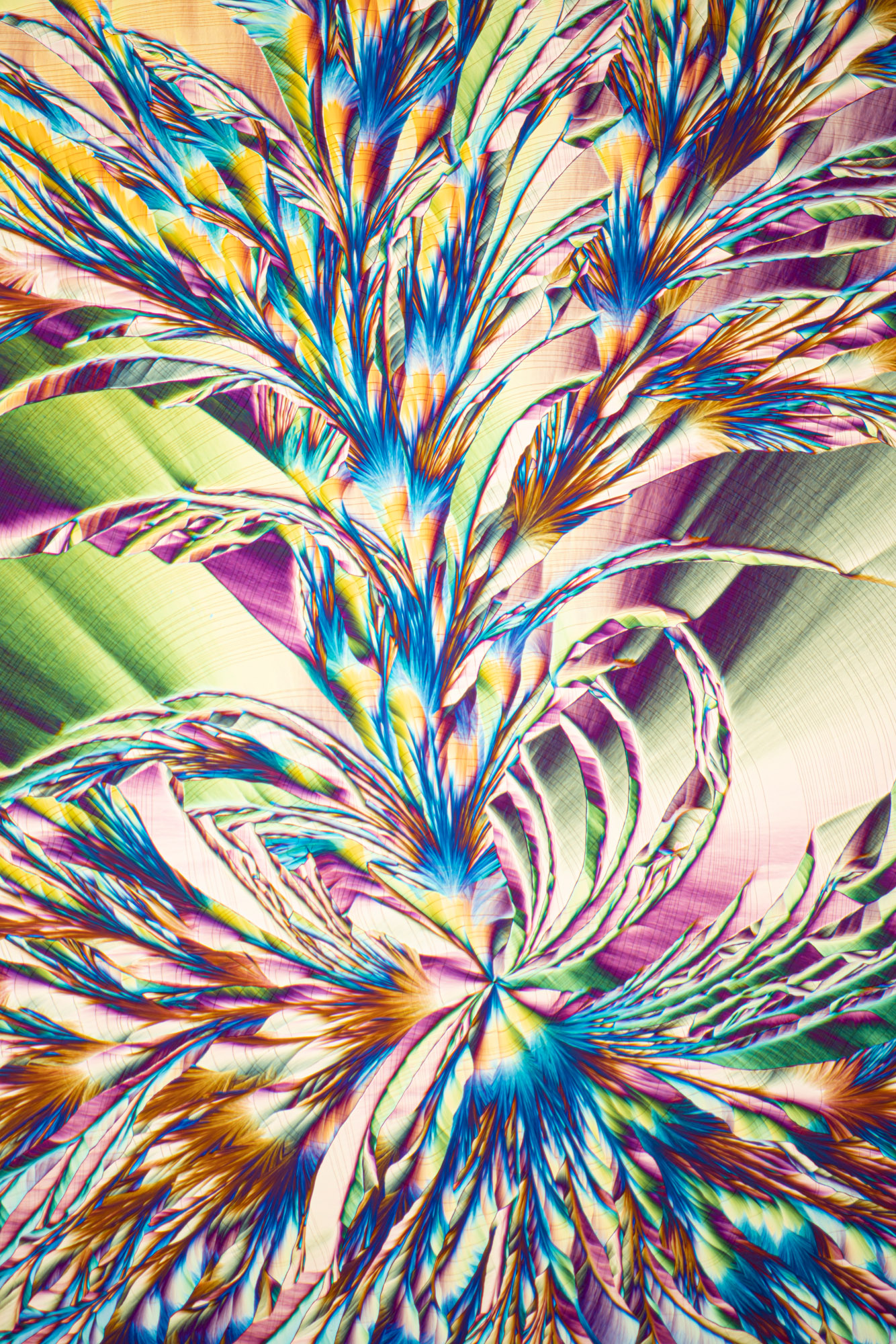
In terms of crystallography, I employ a diverse range of chemicals. This has been a process of research and discovery, keeping a recipe book of the preparation of chemicals for imaging and how the best results are achieved.
Furthermore, I have been mixing chemicals in different ratios to see how the patterns are altered. Sometimes the chemicals, when mixed in a specific ratio, give jaw-dropping results that take on the appearance of plants and flowers.
By adjusting the polarisers and wave plates in the optical train of the microscope, it is sometimes possible to produce a colour palette that makes the scene more like those we see when we wander in a wildflower meadow or a lush deciduous forest in the summertime.
Why are crystals so important to study? What does it mean to you?
Crystals are important to study in terms of deciphering the 3d structure of biological molecules like proteins, in terms of solid-state physics for enhancing technology such as microelectronic devices and increasing the efficiency of solar cells.
As for my work, it is a fusion of science and art. I am motivated by photographing imagery that is both educationally rich and at the same time, aesthetically stunning. I am interested in understanding why the different formations arise, what actually happens when those molecules come together, interact, and how and why they crystallize the way they do.
I would like to understand at a deeper level, how the light interacts with the molecules in the crystal film and how it is manipulated as it passes through.
Do you have a favourite image?
I have several images that I really love and cannot decide which is my favourite.
What are your future ambitions?
I would like to see more of my work published in books that communicate that exquisite beauty of nature on the microscopic scale and the science behind the imagery. To capture slow-motion cinematography of high-speed crystallisation processes and others that form at slow paces but specialised equipment is required for this.
I am hoping at some stage to find a collaborator to produce a series of books with.
Karl's
work in action


