Science Museum
SPL supplied the Science Museum team with images to help contextualise stories in Medicine: The Wellcome Galleries.
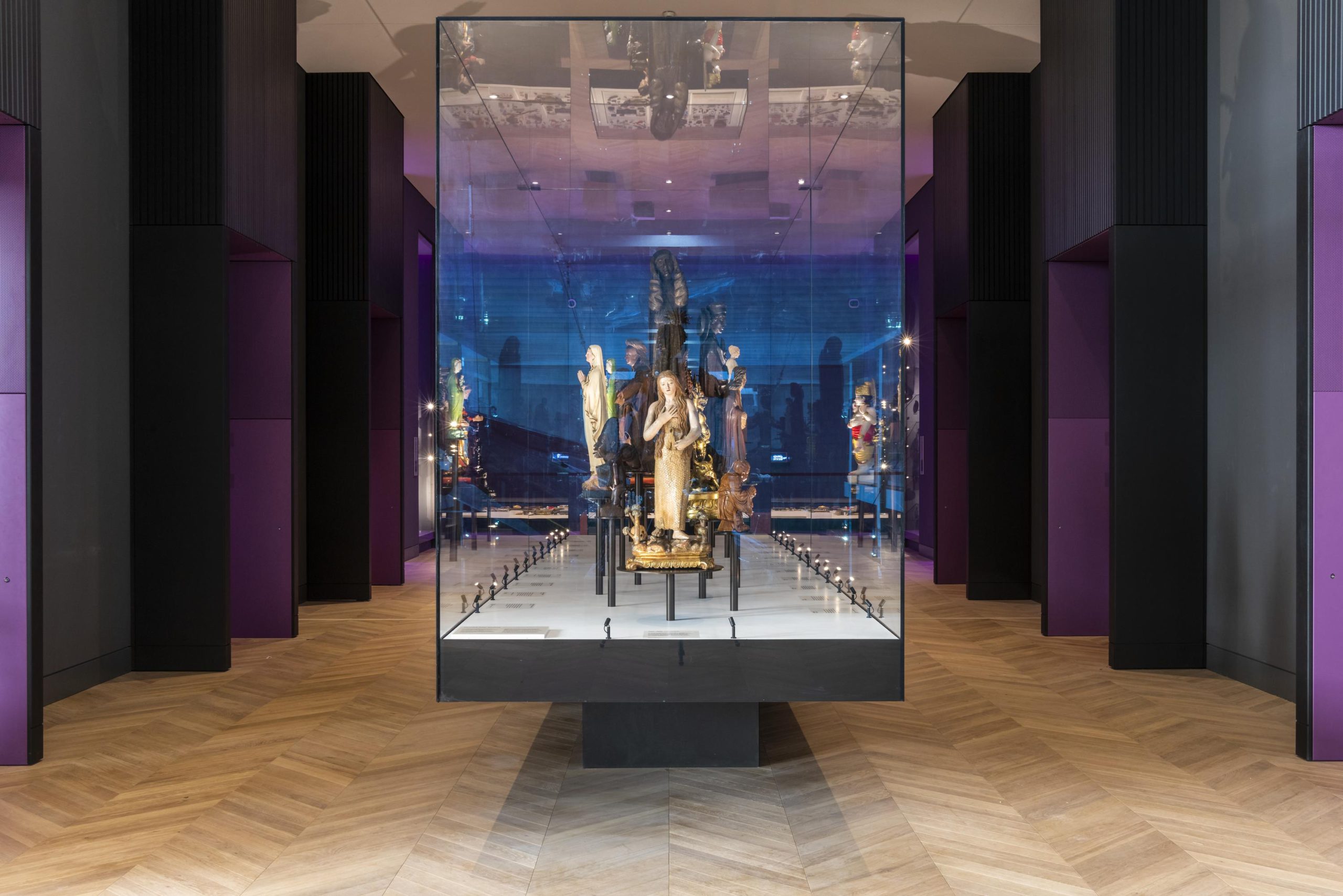
Take a journey covering more than 500 years of medicine. Explore five vast and visually stunning galleries containing more than 3,000 medical artefacts, striking artworks, interactive games and immersive experiences.
Science Photo Library helped show the global public health issue of air pollution and surgical futures using virtual reality.
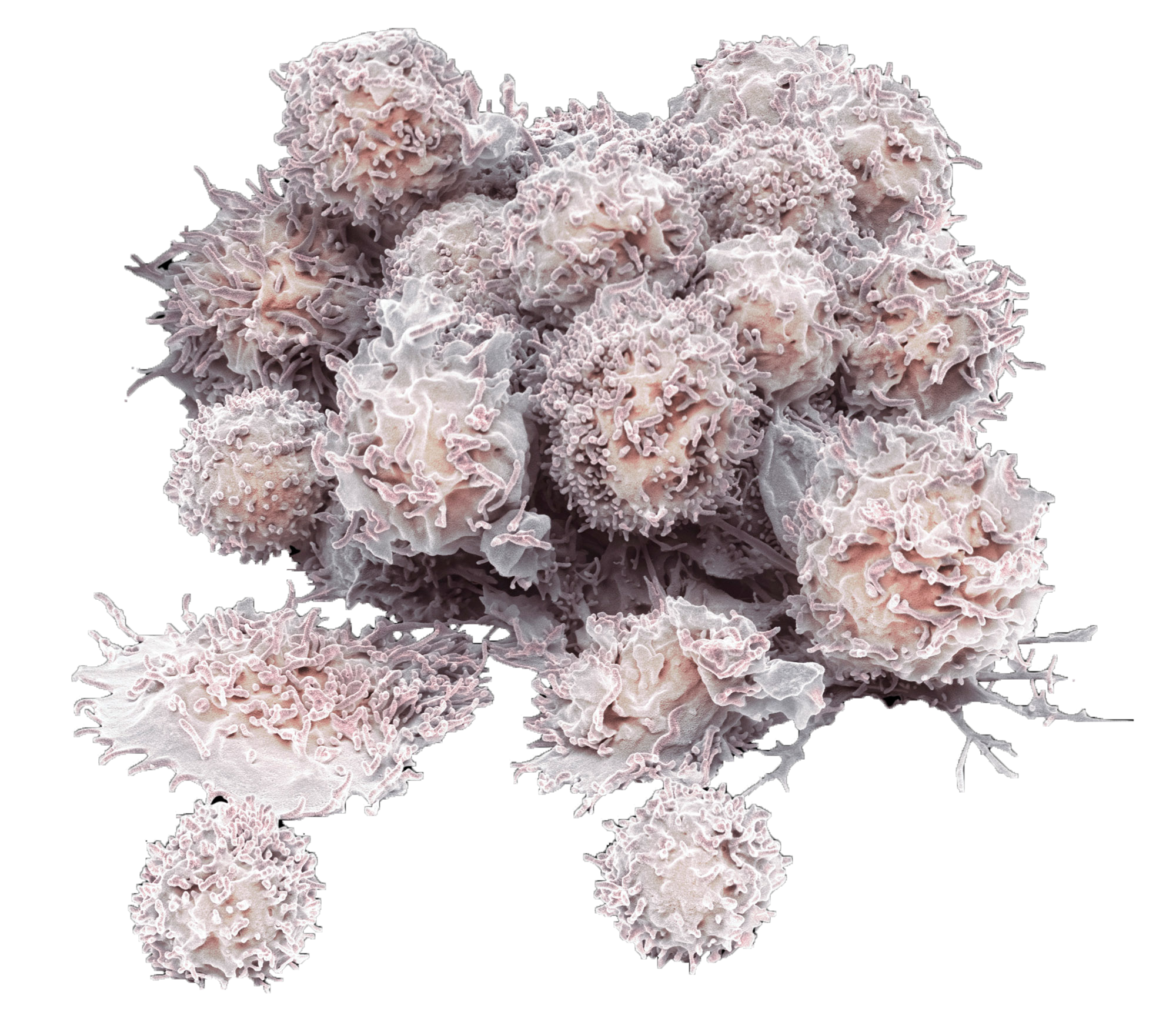
SPL’s images and videos enable visitors to interact with a touch screen table to explore different imaging techniques such as x-ray, ultrasound and MRI. The images are shown in more subtle ways; a magnified view of a cervical cancer cell is the inspiration to create a 3D design for a backdrop focusing on the development of cervical screening.
Project
Exhibit imagery
Client
Science Museum
Science Museum uses our imagery to illustrate medical concepts and enhance visitor curiosity.
The below represents interactive elements of their exhibit.
MRI
Revealing the brain
This sequence of MRI slices shows the internal structure of the head of a patient with a cancerous tumor located in their brain's left frontal lobe. MRI transformed brain imaging by revealing detailed structures inside the brain and how they function. This has made it possible to accurately identify conditions and plan treatment.
MRI
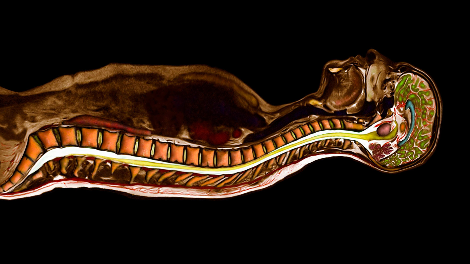
A slice through the spine
MRI is brilliant at imaging differences between body tissues. This image shows a slice through the centre of the body revealing the brain, chest cavity, and spine. MRI images are usually black and white, but here the images have been coloured. The brainstem (yellow) becomes the spinal cord (yellow), which is encased by the vertebrae (orange) of the spine.
See our collection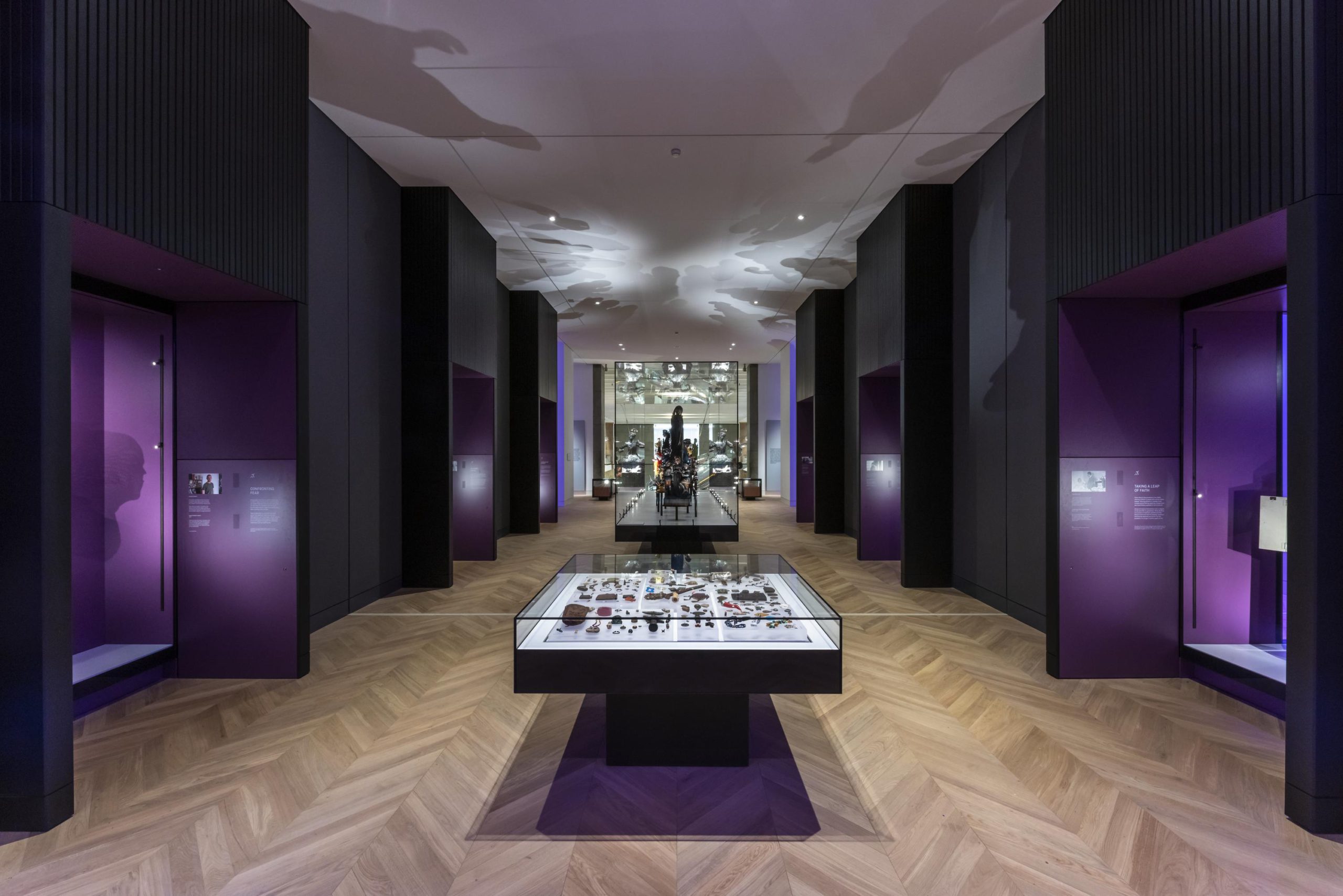
‘The vast array of image and film available made Science Photo Library one of the go to places to look for supporting material to bring stories on the topic of medicine over the last 500 years to life.’
Keeley Carter
Client Project Manager
Science Museum


")

