Vsevolod Zviryk
Views from within
He began to experiment with different perspectives and colours, adding a creative spark to his professional work. At the moment, his country is facing challenging war time conditions, but Vsevolod still manages to create stunning content.
Tell us about your background. How did you become interested in medicine?
I was born into a family of medical professionals. My father was an X-ray technician and my mother was a nurse, so practicing medicine was a natural choice.
I knew that I would become a doctor since I was in junior high school. I liked the humanities the most – history, geography and biology. At the same time, I was always interested in electronics and photography.
I started to understand the circuitry of electronic devices on my own, learned to solder and to repair household or computer equipment. Therefore, children’s hobbies and interests influenced my subsequent choice of specialisation; radiology is a combination of medicine and electronics.
Where did you study and did you specialise in a particular medical field?
After school, I studied for four years at the local Chortkiv medical college, where I mastered basic medical skills and worked as a doctor’s assistant for a while.
I then studied at the Ivano-Frankivsk Medical University and after 6 years of study and 2 years of internship, I received my first specialisation in general medical practice.
I received my second specialisation in radiology two years later in Kyiv.
What led you to medical imaging?
At a lecture on radiology I saw a simple 3D visualisation of a CT examination for the first time. It made an impression on me and the same day at home I started looking for information and software for independent processing of DICOM images. After a few hours, I managed to get a three-dimensional image.

When did you come up with the idea of sharing your work with the public?
Almost immediately. When I heard favourable reviews from my colleagues, I started posting my work in various social networks and forums.
How did your collaboration with SPL start?
Seymour Yang from SPL wrote to me and offered to place my work with SPL. I agreed.
Soon after I got to know Rose Taylor, who selected the most suitable images and videos.
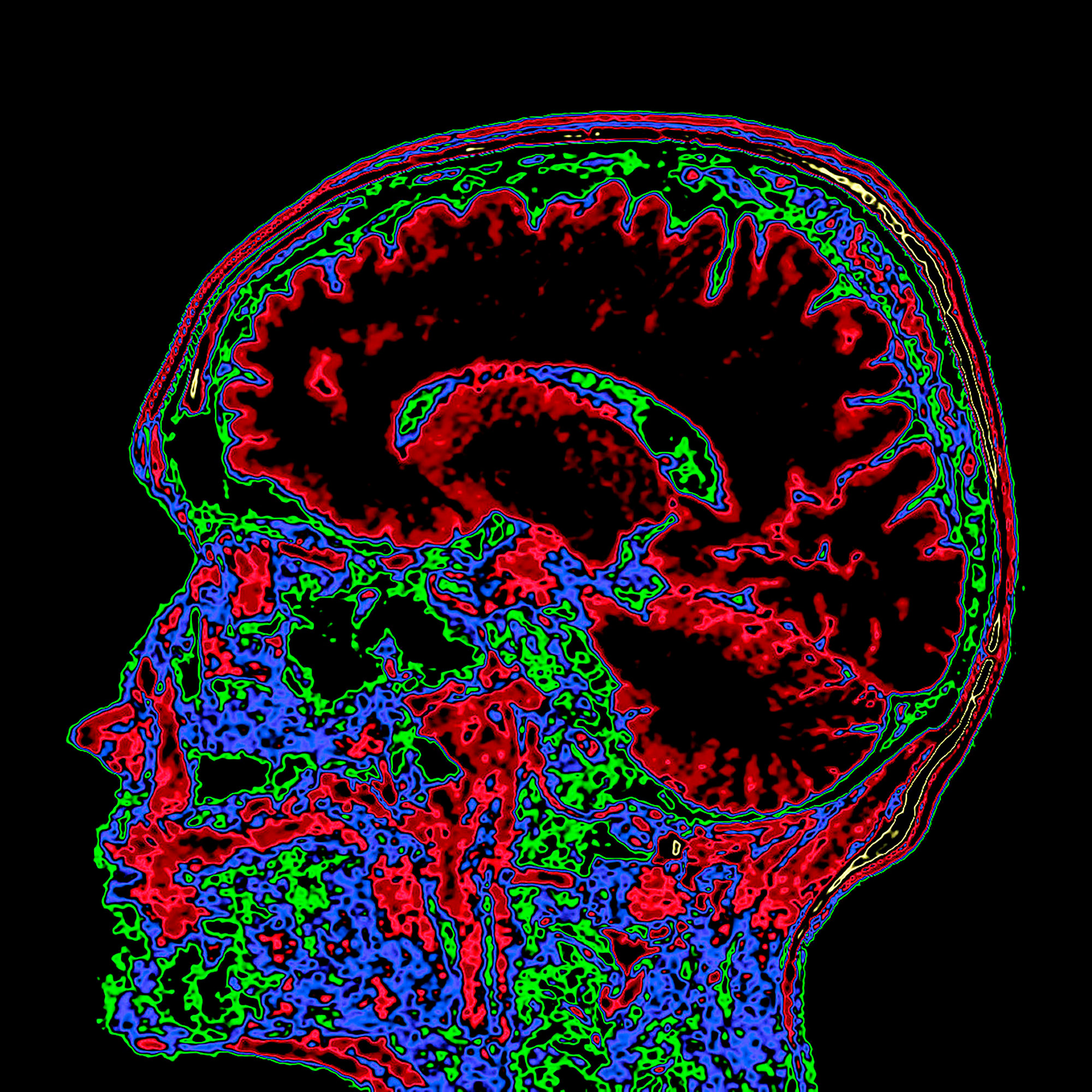
What inspired you to give your diagnostic images an artistic twist?
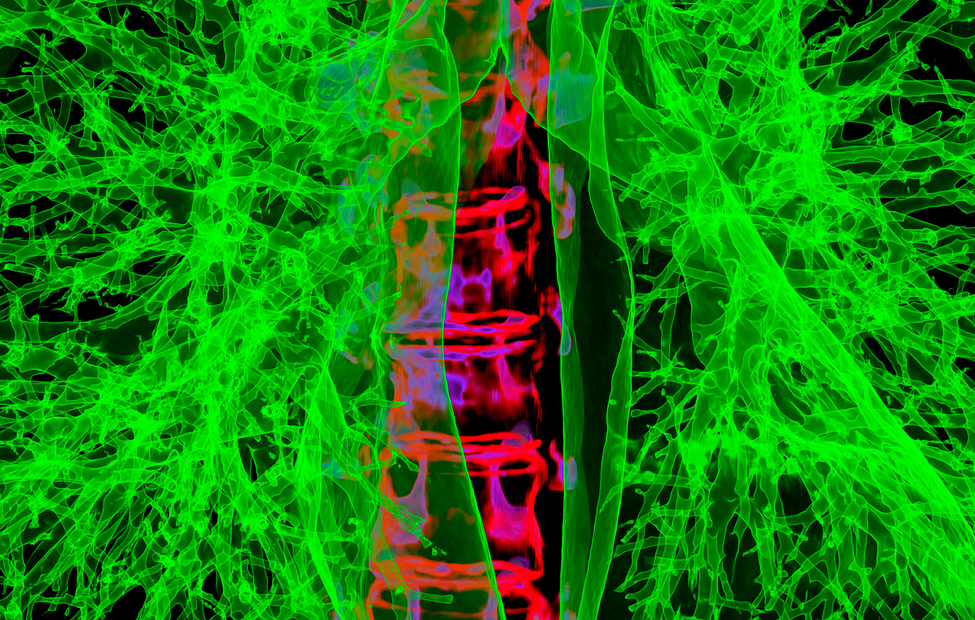
Most of the time, radiologists work with black and white images.
A multi-coloured, bright image is a way to shift the gaze and perceive things differently.
Also, I like to experiment with different photo filters and settings.
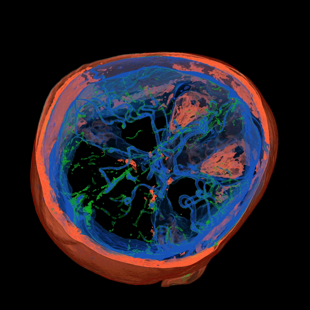
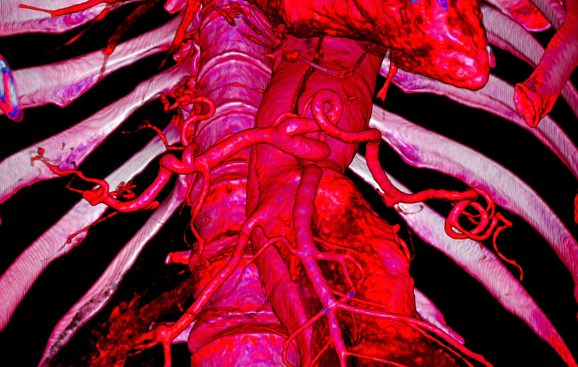
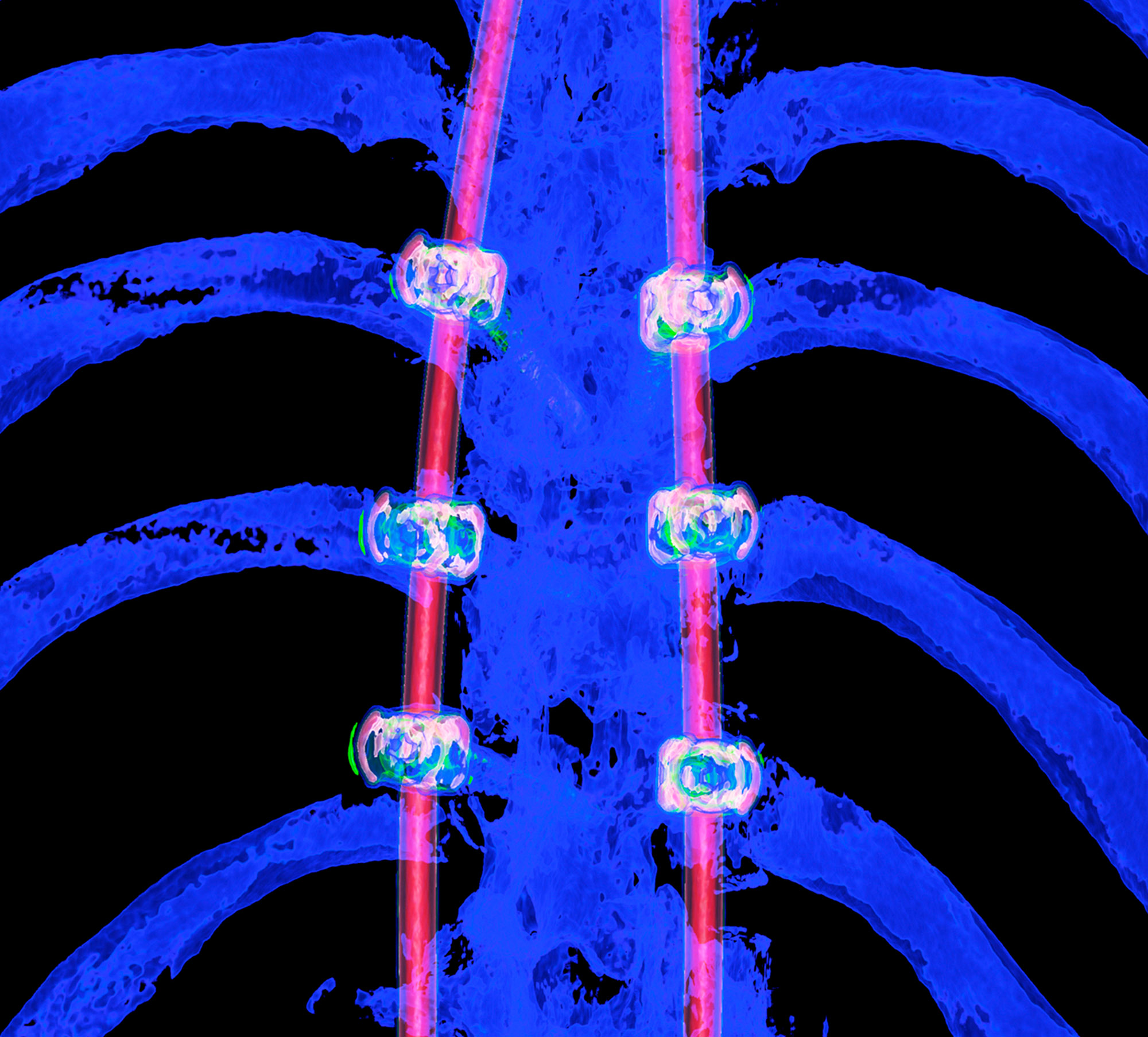
We love some of the extraordinary angles you get when producing an image? How do you do it?
The program for processing radiological images (DICOM archives) allows you to create and look at a three-dimensional image from different angles.
If you change the location of the virtual lens, thereby changing the perspective, you can get such images.
How do you decide which colours to use on the images?
The colours choose themselves.
I simply create a table where the radiological density of the material can correspond to any colour.
After combining different shades, I only have to choose what I like the most.
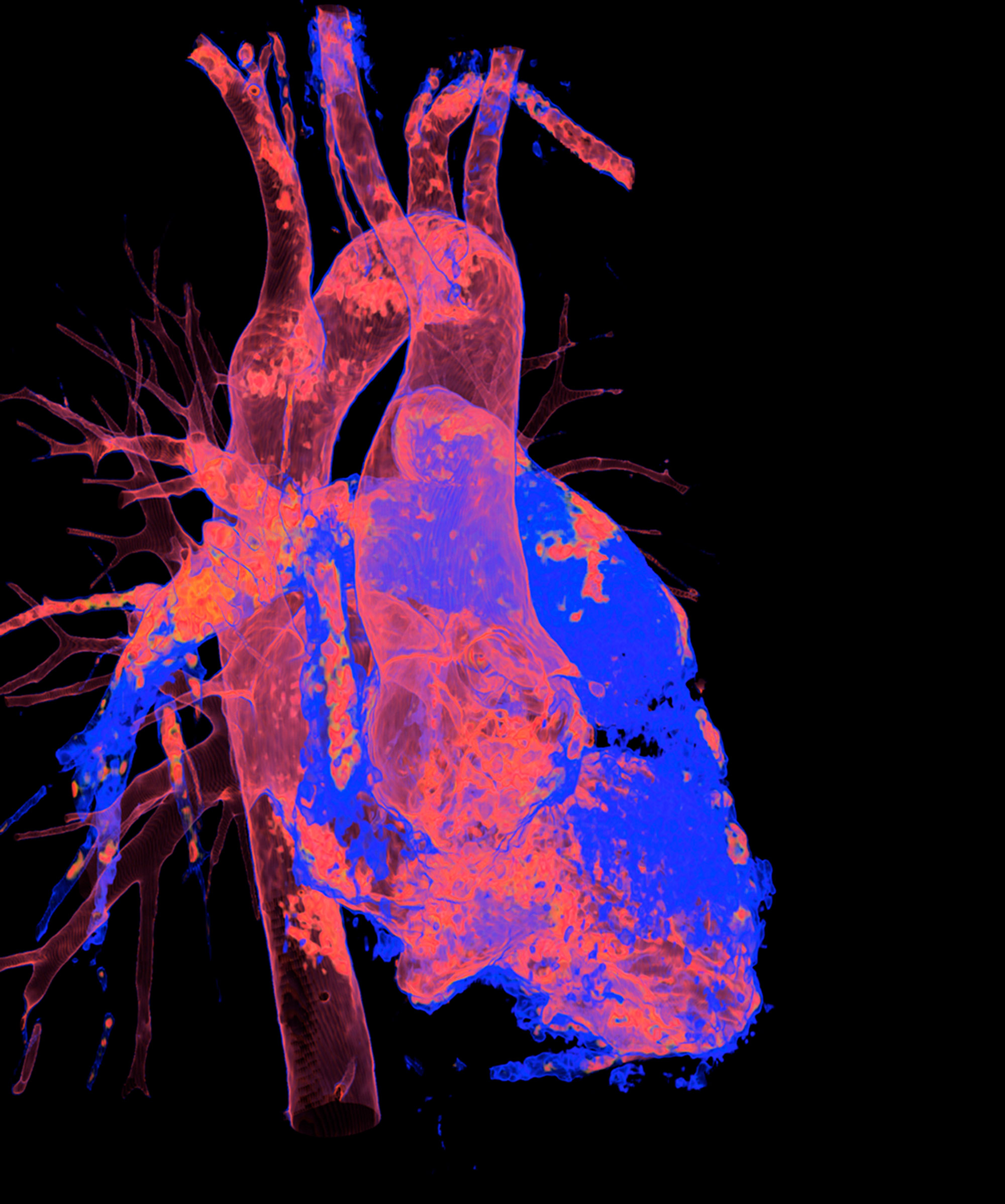
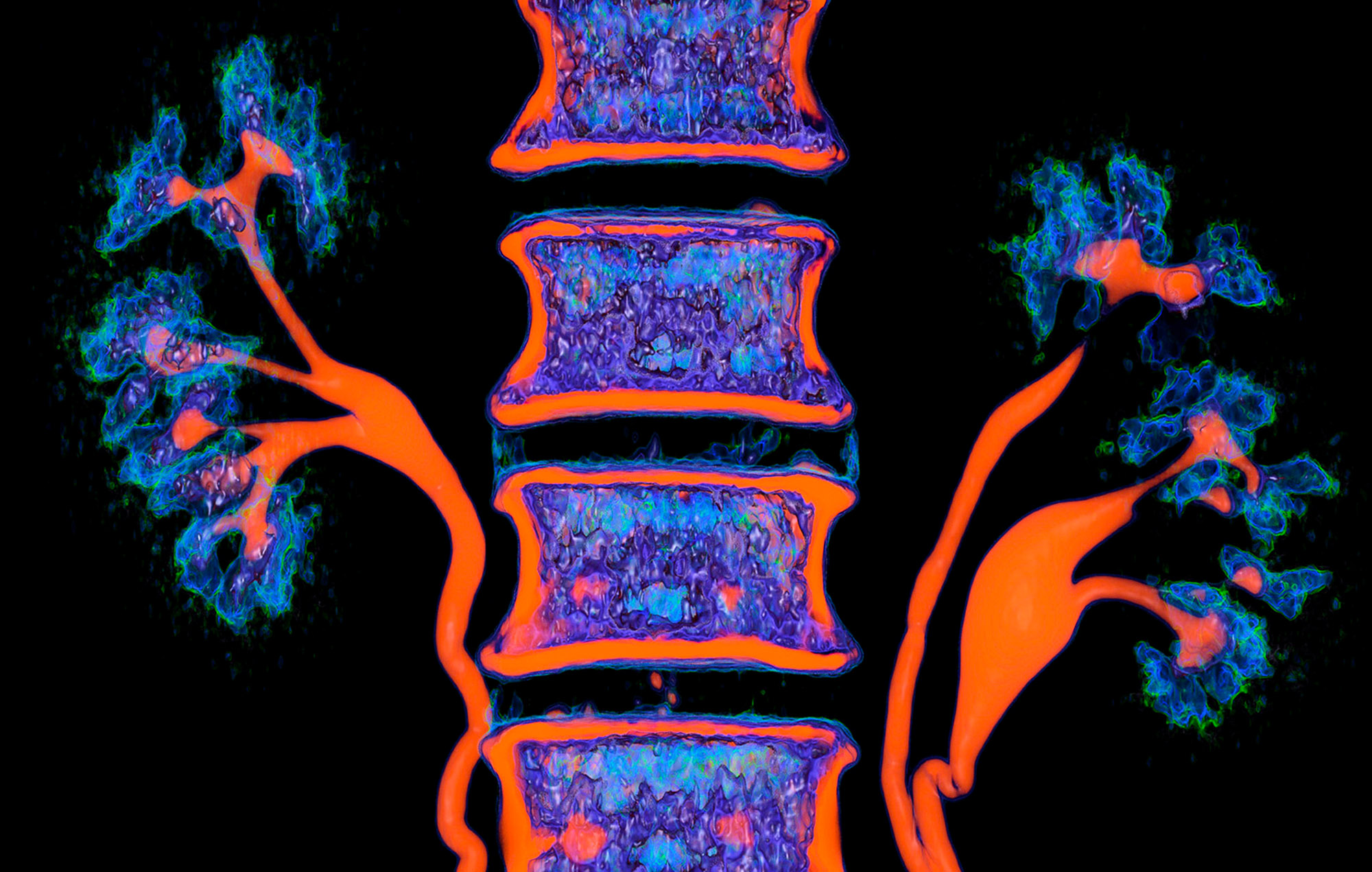
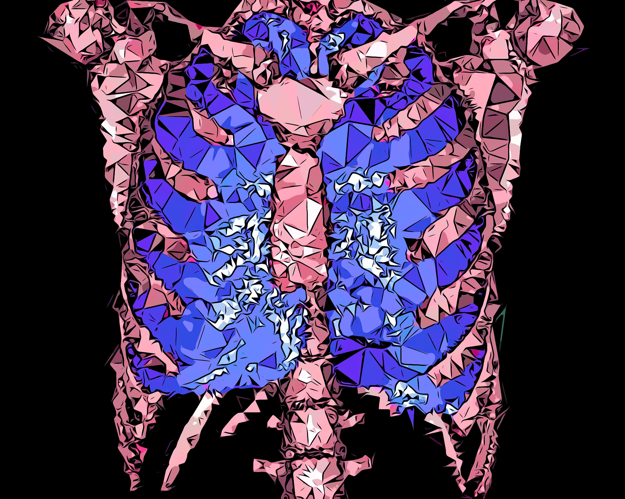
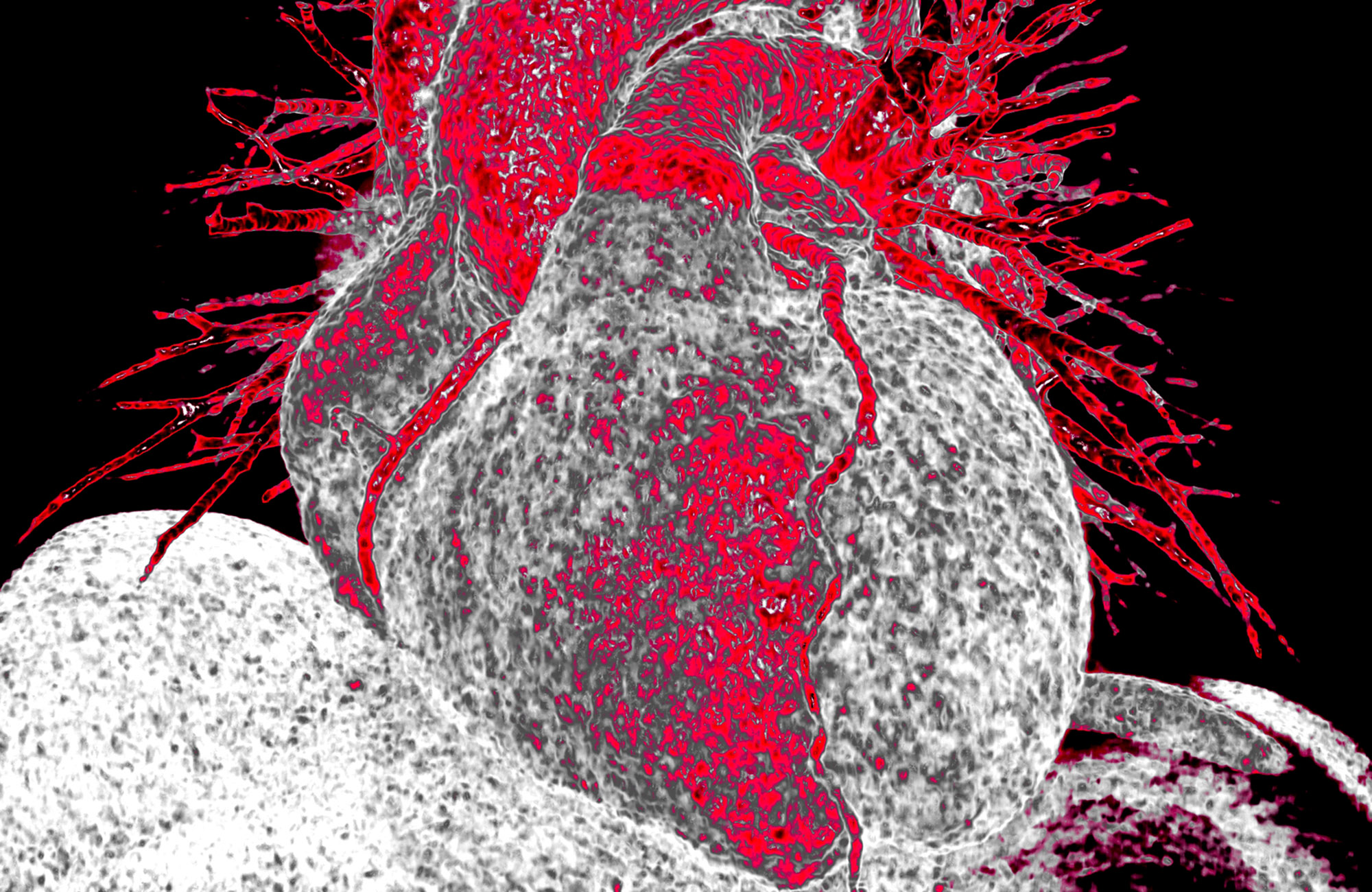
Is there a particular area of the body you specialise in?
No. Tissues with different densities are best suited to visualisation by CT imaging.
Lungs and heart, bones and muscles or organs injected with a contrast agent reveal the best structure and anatomy.
Where and how would you like to see your images used?
I really like to see my work in specialised medical literature.
I am especially happy when my work gets on the cover of a publication or is used to advertise a medical event.


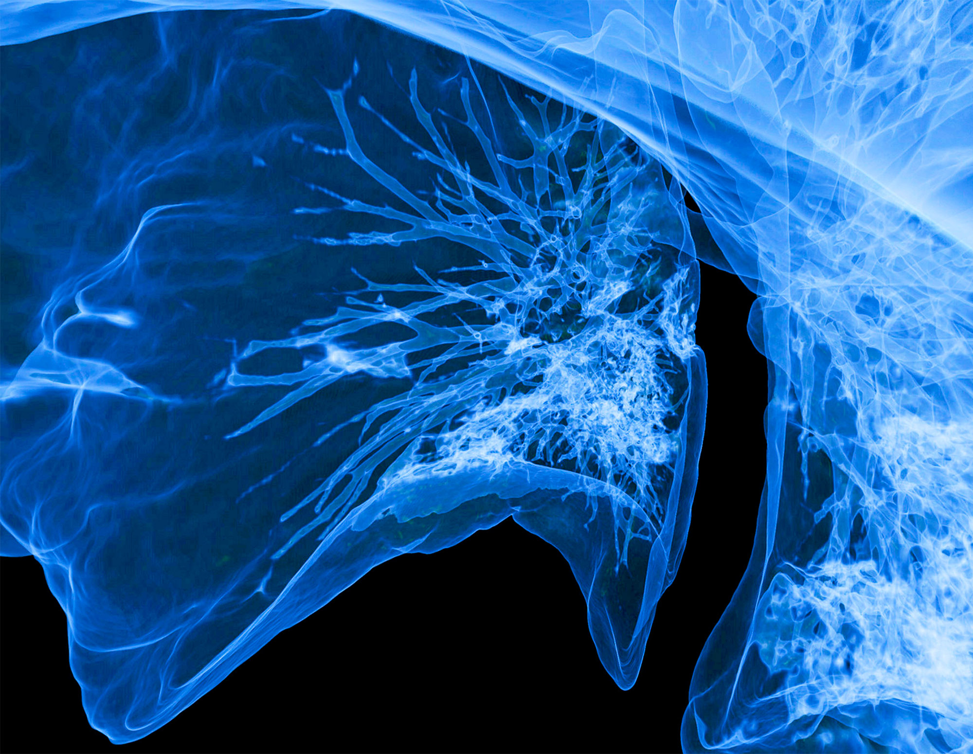
Do you have a favourite image and what makes it special to you?
My favourite image is a 3D CT scan of lung airways.
It was one of my first images using a completely redesigned tissue density colour table. Green is the most dense tissue (bone) and purple is the least dense tissue (lung).
Combining these colours resulted in an extremely bright artistic image.


